The Aberrant Right Subclavian Artery in Normal Fetuses
Vladimir Lemaire (1), Shivani Raman Patel (1), Ashley Zink (1), Gabriella Ricciardi (2), Patricia Santiago-Muñoz (1), Robyn Horsager-Boehrer (1)(1) UT Southwestern Medical Center, (2) CHU de Lille
Article Published: Aug 25, 2023
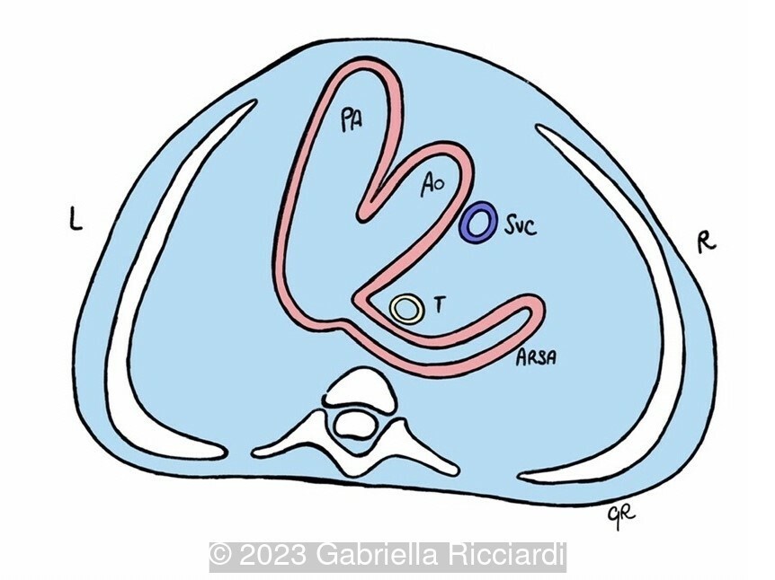
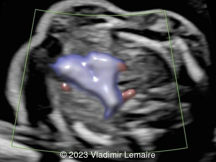
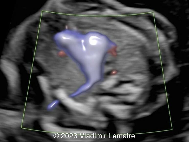
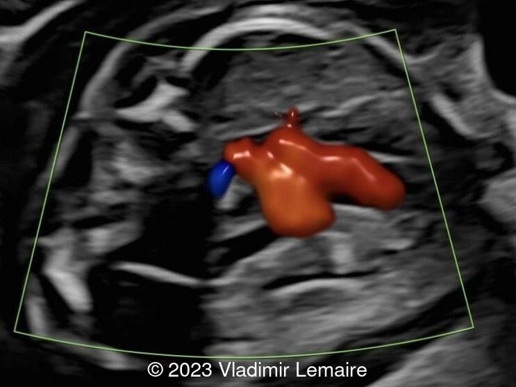
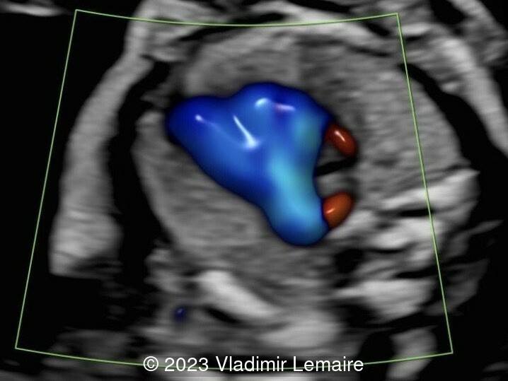
The aberrant right subclavian artery, also called retroesophageal right subclavian artery or Lusoria artery, results from partial regression of the right aortic arch between the right common carotid artery and the right subclavian artery. Consequently, a left-sided aortic arch persists, giving rise to the vessels in the following order: the right common carotid artery, the left common carotid artery, the left subclavian artery, and the aberrant right subclavian artery (ARSA).
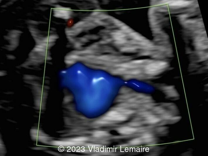
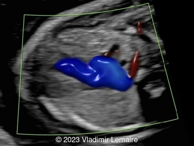
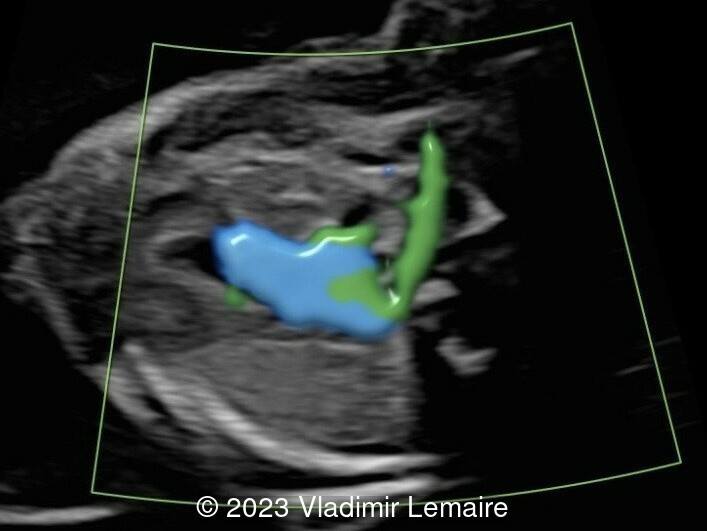
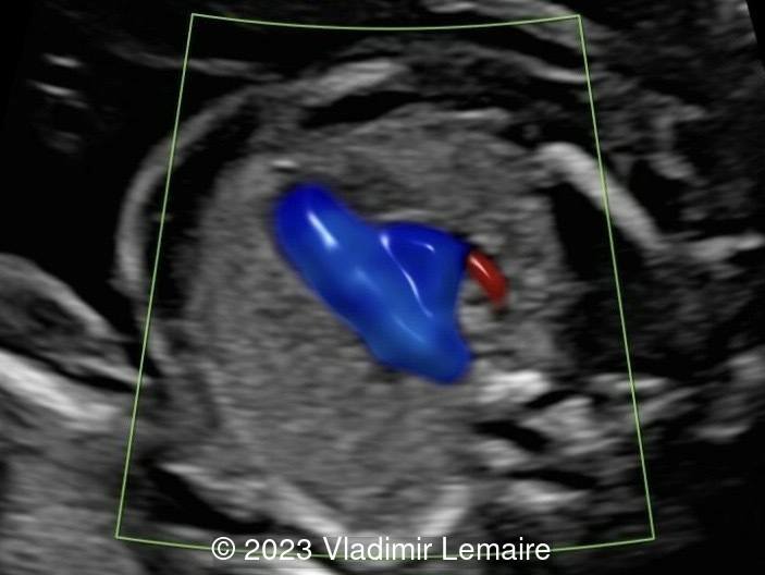
A left-sided aortic arch with an ARSA is the most common aortic abnormality, occurring in up to 1.5% of the general population. Typically visualized when color Doppler is applied at the level of the three-vessel-trachea view, it can be demonstrated emerging from the junction of the aortic arch and the ductus arteriosus, coursing behind the trachea and esophagus toward the right arm.
An ARSA can be associated with conotruncal anomalies, which increases the risk for microdeletion 22q11.2. It can also be found in aneuploidies such as trisomy 21, trisomy 18, trisomy 13, Turner syndrome, deletion 4p-, etc.
When isolated, as reported by Zalel et al., an ARSA can be considered a normal variant. For normal fetuses with low risk noninvasive prenatal testing, there is no need for invasive prenatal testing.
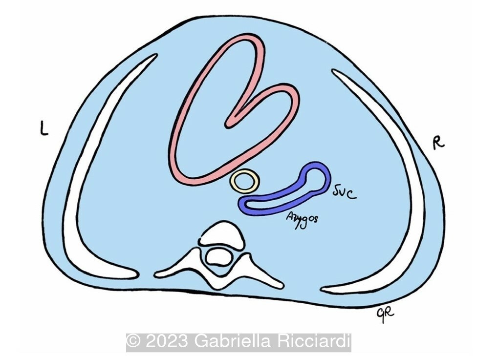
The azygos vein, which courses on the right side of the spine and trachea before it drains into the superior vena cava (SVC), can be confused with an ARSA. In case of doubt, an arterial Doppler waveform, when pulsed wave Doppler is applied, can help differentiate an ARSA from the azygos vein.
Complications due to compression of the esophagus by an ARSA (dysphagia lusoria) are rare.
References
[1] Abuhamad, A, et al. "Aberrant Right Subclavian Artery". A Practical Guide to Fetal Echocardiography: Normal and Abnormal Hearts (4th edition). Singapore: Wolters Kluwer, 2022. pgs 599-604.
[2] Zalel, Y, et al. Fetal aberrant right subclavian artery in normal and Down syndrome fetuses. Ultrasound Obstet Gynecol. 2008; 31: 25-29.