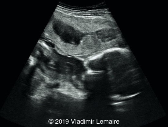
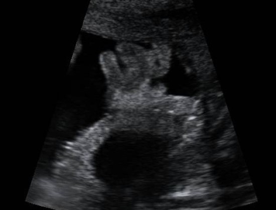
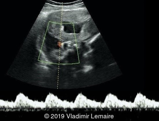
Placental Abruption with retroplacental hematoma
Vladimir LemaireMaternite Rita Merli, Hopital Saint Damien, Nos Petits Freres et Soeurs, Haiti
Article Published: Dec 20, 2018
Placental abruption is the detachment of a normally inserted placenta that occurs in the third trimester of pregnancy, usually in the setting of pregnancy-induced hypertension such as pre-eclampsia. The hematoma can be retroplacental, subchorionic, and less commonly, preplacental.
The diagnosis of placental abruption is primarily based on clinical signs and symptoms such as abdominal pain and vaginal bleeding. Ultrasound can be used in the diagnosis, although its sensitivity remains poor with only 25 to 50% of cases detected by ultrasound. Findings on ultrasound include a hyperechoic, isoechoic, or hypoechoic (as presented in the present case) collection of blood between the uterine wall and the placenta.
If the pregnancy is at term or near term, it is best to deliver the patient. Before 34 weeks of gestation, a conservative approach with strict maternal and fetal monitoring can be adopted if the maternal condition is stable and the abruption not expanding in order to induce fetal lung maturation.


References
[1] Doubilet P, et al. "Placenta." Atlas of Ultrasound in Obstetrics and Gynecology, A Multimedia Reference. Philadelphia: Wolters Kluwer, 2019. pgs 311-312.
[2] Campbell W. "Placental Abruption in a Twin Pregnancy." TheFetus.net. https://thefetus.net/content/placental-abruption-in-a-twin-pregnancy, Publish date 05/2002.
[3] Sotiriadis A, et al. ISUOG Practice Guidelines: role of ultrasound in screening for and follow-up of pre-eclampsia. Ultrasound Obstet Gynecol. 2019;53(1):7-22.