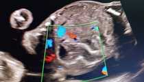
Fetal hepatic SOL at 21 wks POG with altered maternal LFT's
Fetal hepatic SOL at 21 wks POG with altered maternal LFT's
#1 -
01/01/2022
said
“Hi Deepshikha,
Thank you for sharing of your case. I think that prenatal picture of hepatic masses is never specific enough to be completely sure about correct diagnosis. Seeing the images, the findings could represent liver hemangioma or hepatoblastoma. I would repeat the examination every 2 or 3 weeks to see progression or regression of the lesion and I would search for some possible signs of cardiac failure, hydrops, polyhydramnios, etc. If it is possible, fetal MRI should be offered to the patient. Also, amniocentesis should be done to search for possible association with BWS (Beckwith–Wiedemann Syndrome) especially if there are some other signs of fetal organomegaly. I would wait until term or until signs of cardiac failure occur and then deliver the baby. Final diagnosis and management will depend on postnatal findings.
Franti”
#2 -
01/02/2022
said
“Dear Deepshikha .A) Your presentation is inappropriate. We need slow video clips axial and sagittal sections , we need good calibrated colored flow in the lesion (to exclude Caroli disease ).I don't understand the relation between the hypoechogenic semi round mass inside the liver and the elongated with hyperechogenic borders "loop" in the level of the intestines.Is it the same or two different entities .
B) There is a long DD of hepatic mass from hematoma secondary to CMV to benign tumor the most frequent hemangioma to malignant tumor like hepatoblastoma .
C) I have one case of maternal severe jaundice secondary to autoimmune response to drug and fetal hepatomegaly. The fetal recovery was simultaneous to maternal recovery the child is doing well.”
#3 -
01/04/2022
said
“Thanks ”
#4 -
01/04/2022
said
“Thanks for valuable opinions.
@ Moshe, I will take the clips and post in next visit.
Linear echogenic foci along the hepatic borders, is echogenic bowel.
”
@ Moshe, I will take the clips and post in next visit.
Linear echogenic foci along the hepatic borders, is echogenic bowel.
”
#5 -
01/21/2022
said
“Maternal health progression will probably dictate course of care for next two months. If stable then fetal well being after 28 weeks will guide action regarding pregnancy. In my humble opinion.”
#6 -
01/22/2022
said
“Hello everyone
I want to tell followup of above mentioned case.
Mother had cervical incompetence with dilatation at 24 weeks. Baby was delivered within few hrs without steroid coverage.
Was in NICu for 2days. AFP done was >1000ng/ml.
Post mortem biopsy of hepatic Sol showed hepatoblastoma with low mitotic activity n osteoid component.
Now only question is , how much will be chances of recurrence in subsequent pregnancy?”
#7 -
01/22/2022
said
“Is that the internal os fully opened in the view of transverse abdomen? ”


Needed opinion about a case.
A primi of 21wks POG. fetus was diagnosed with Well defined, irregular marginated hypoechoeic avascular hepatic SOL with mild polyhydroaminos n non visualised ductus venosus.
Mother has severe itching n altered SGOT n SGPT
Now how to proceed further in this scenario?
Regards