Case of the Week # 661
(1) Azienda Sanitaria Provinciale di Cosenza, Cosenza, Italy; (2) Centro Diagnostico Di Meglio, Naples, Italy; (3) Presidio Ospedaliero "Giovanni Paolo II", Lamezia Terme, Catanzaro, Italy; (4) Laboratorio di Genetica Medica, Ospedale Pediatrico Bambino Gesù, IRCCS, Rome, Italy; (5) Liverpool Women's NHS Foundation Trust, Liverpool, United Kingdom; (6) Department of Genetics, Polish Mother’s Memorial Hospital – Research Institute, Łódź, Poland
A 43-year-old nulliparous woman with an unremarkable personal and family history conceived spontaneously. First-trimester screening performed elsewhere was reported as normal. Non-invasive prenatal testing for trisomies 21, 18, and 13 was low risk (fetal fraction 13%). At 21 weeks and 3 days, an abnormal second-trimester anomaly scan prompted detailed fetal ultrasound assessment. The following findings were identiifed. What is the most likely diagnosis?
View the Answer Hide the Answer
Answer
We present a case of Raine syndrome (FAM20C-related osteosclerotic dysplasia).
After review of the imaging, the key prenatal findings were:
- Abnormal cranial contour with a trigonocephalic configuration (trigonocephaly) and premature metopic suture closure (metopic synostosis)
- Marked bilateral periventricular hyperechogenicity, subsequently shown to represent parenchymal calcification (periventricular calcification, HP:0007229*)
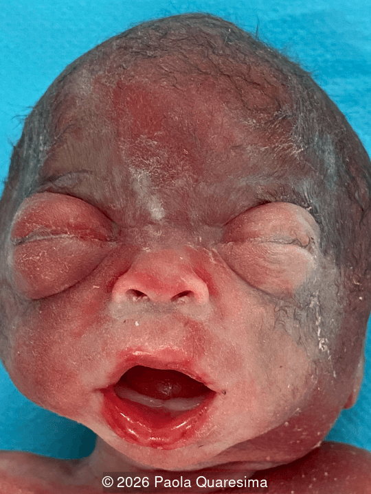
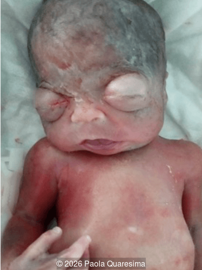
- Hypertelorism and extreme ocular proptosis/exophthalmos (HP:0000520*)
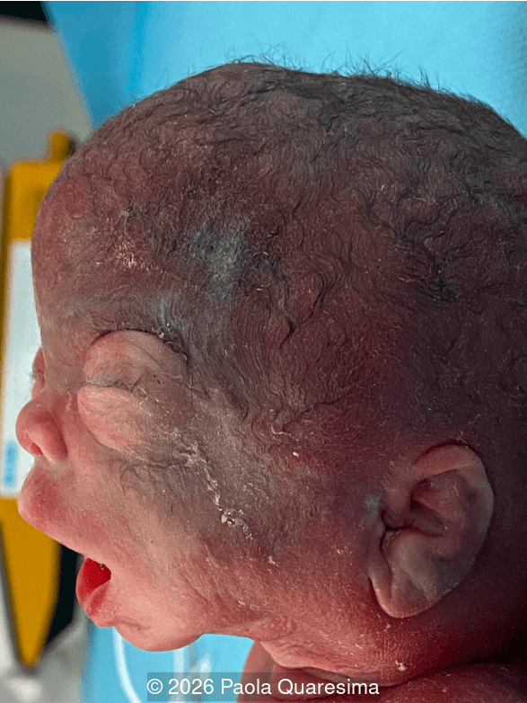
- Midface and lower-face hypoplasia (midface retrusion, HP:0011800*) with an abnormal retrognathic facial profile (micrognathia / retrognathia)
- Increased nuchal fold
- Prematurely visible eyelashes and prominent scalp hair for 21 weeks of gestation.
The combination of intracranial mineralization with a distinctive craniofacial phenotype is highly suggestive of Raine syndrome.
Second- and third-opinion ultrasound examinations confirmed marked periventricular cerebral hyperechogenicity and facial dysmorphism. Amniocentesis was performed. Chromosomal microarray (molecular karyotype) and clinical exome sequencing were initially reported as normal/negative. Fetal brain MRI was also undertaken and reported as unremarkable.
Following multidisciplinary counselling, the parents opted for termination of pregnancy. Postmortem examination confirmed severe proptosis, trigonocephaly with marked metopic ridging, hypoplastic nose with depressed nasal bridge, tented "fish-like" mouth, high-arched palate, gingival prominence, and conspicuously developed eyelashes and scalp hair.
Because the clinical phenotype remained pathognomonic for Raine syndrome, a fetal skin biopsy was submitted for advanced molecular investigation. While targeted screening for recurrent single-nucleotide variants was negative, extended genomic analysis incorporating Whole-Genome Sequencing (WGS) and high-resolution copy-number variation (CNV) unmasked the underlying compound heterozygosity in FAM20C (chromosome 7p22.3):
- A paternally inherited microdeletion encompassing exon 3 of FAM20C;
- A complex structural rearrangement involving an inversion/translocation breakpoint (genomic coordinates chr7:200786–503085, GRCh38) disrupting the FAM20C locus, confirmed by Sanger sequencing.
Histological examination of the fetal periventricular brain parenchyma demonstrated numerous dystrophic calcifications, confirming the pathological basis of the sonographic hyperechogenicity.
*The Human Phenotype Ontology (HPO) provides a standardized system in which individual phenotypic features, postnatal or prenatal, are assigned unique, unambiguous codes. This is particularly valuable in fetal medicine, where the same ultrasound finding may be described using different terms or synonyms. HPO coding facilitates precise communication between ultrasound specialists, clinical geneticists, and molecular laboratories and allows the fetal phenotype to be systematically matched with genomic findings. Accurate phenotypic coding can therefore improve variant interpretation, genotype-phenotype correlation, and ultimately diagnostic precision while reducing ambiguity arising from differences in terminology.
Discussion
Raine syndrome (OMIM #259775) is a rare autosomal recessive disorder caused by biallelic pathogenic variants in FAM20C on chromosome 7p22.3 [1,2]. FAM20C encodes a Golgi-associated kinase that phosphorylates serine residues within S-X-E/pS motifs across secretome proteins, playing an indispensable role in biomineralization, extracellular matrix organization, and systemic phosphate homeostasis [2,4]. Although classically regarded as an invariably lethal neonatal osteosclerotic bone dysplasia, the recognized phenotypic spectrum extends from lethal neonatal forms to attenuated, non-lethal forms with survival into adulthood [2,3].
The classic phenotype encompasses generalized osteosclerosis, cranial hyperostosis, intracranial calcifications, and distinctive craniofacial dysmorphism [1,5]. Characteristic features include exophthalmos/proptosis, hypertelorism, midface hypoplasia, depressed nasal bridge, micrognathia, a small tented mouth ("fish-like" facies with tented upper lip vermilion), gingival hyperplasia, and a cleft palate or high-arched palate [2,5,6]. Severe neonatal presentations frequently feature thoracic hypoplasia and lethal pulmonary hypoplasia [2]. Not all skeletal manifestations (such as marked long-bone hyperostosis) are prominent during the second trimester, making cranial and facial signs paramount for early detection [1,6].
The Brain: Raine Syndrome mimics fetal infection
The cerebral appearance is an important diagnostic trap because it initially mimics congenital intrauterine infection, particularly cytomegalovirus (CMV). In routine fetal neurosonography, marked periventricular hyperechogenicity instinctively points to congenital TORCH infections, predominantly cytomegalovirus (CMV) or toxoplasmosis [7,11]. Periventricular calcifications in CMV typically reflect necrotizing ependymitis and calcified subependymal glia, coexisting with ventriculomegaly (lateral ventricle dilatation), subependymal cysts, temporal lobe cysts, or cortical dysplasias [7].
Raine syndrome belongs to an essential group of genetic mimics of congenital infection ("pseudo-TORCH" phenotypes) [11]. The mineralization in Raine syndrome selectively affects the ventricular margins, basal ganglia, and pericallosal tissue [1,3]. Therefore, neurosonographic topography alone overlaps substantially with CMV.
Extracerebral facial morphology resolves this ambiguity. Severe proptosis, metopic craniosynostosis/trigonocephaly, midface retrusion, and premature hair/eyelash development are absent in congenital CMV. When periventricular calcification coincides with a craniofacial dysplasia pattern, non-infectious genetic etiologies must be considered [1,5].
Proptosis as a critical second diagnostic axis
A systematic diagnostic strategy requires assessing the neuroaxis and the craniofacial profile along independent diagnostic vectors:
- When viewing the brain alone, periventricular calcification suggests CMV or a pseudo-TORCH phenotype [7,10,11].
- When viewing the orbits and calvarium alone, proptosis and trigonocephaly suggest syndromic craniosynostosis (FGFR2/FGFR3 spectrum) [5].
Neither model alone is sufficient. The intersection of intracranial calcification with ocular exorbitism/proptosis and facial hypoplasia defines Raine syndrome [1,5,6]. Metopic suture involvement and trigonocephaly can occur secondary to the aberrant mineralization kinetics induced by FAM20C deficiency [5].
Eyelashes in the fetus
Prominent eyelashes is an unusual finding at this early stage. Although potentially representing a minor dysmorphic feature, prominent eyelashes may also reflect familial variation and are not an established characteristic of Raine syndrome. Their appearance may broaden the differential diagnosis to conditions associated with eyelash trichomegaly or hypertrichosis, particularly Cornelia de Lange syndrome and, less specifically, Kabuki syndrome. This observation should therefore be interpreted within the overall fetal phenotype rather than regarded as a syndrome-specific marker. Additionally, nonsyndromic, transient fetal hypertrichosis has also been reported.
The diagnostic limitation of fetal MRI
The unremarkable fetal MRI in this case provides a crucial radiological insight. While MRI provides superior soft-tissue contrast for gyration, opercularization, and white-matter myelination, it is frequently insensitive to subtle, punctate mineral deposits [12]. Calcium demonstrates high acoustic impedance, rendering minuscule subependymal calcifications strikingly hyperechoic on ultrasound long before they generate significant T1-shortening or susceptibility artifacts on conventional fetal MRI [1,12]. Therefore, a negative fetal MRI should never discount distinct calcifications visualized on high-frequency neurosonography.
Genetic testing pitfalls: navigating negative exome sequencing
In this case, routine clinical exome sequencing was initially negative, as single-exon deletions and non-coding structural breakpoints are frequently missed by standard short-read exome capture pipelines. A negative clinical exome does not rule out monogenic disease when the ultrasound phenotype is compelling.
- Coverage and capture gaps: Short-read next-generation sequencing can underperform in GC-rich regulatory domains, deep intronic regions, and repetitive sequences [6].
- Variant classes: Exome bioinformatic pipelines optimize for coding SNVs and small indels. Single-exon deletions/duplications (such as the paternal FAM20C exon 3 deletion in this fetus) require dedicated CNV callers or array-CGH [6]. Non-coding structural variants (inversions, balanced translocations) typically require Whole-Genome Sequencing (WGS) or optical genome mapping.
- Aneuploidy screening: Cell-free DNA (NIPT) screens exclusively for common aneuploidies and does not assess rare autosomal recessive conditions.
Differential diagnosis
Entity | Primary Cerebral Findings | Craniofacial / Extracerebral Clues | Distinctive Discriminator from Raine Syndrome |
Congenital CMV [7] |
|
|
|
Congenital Toxoplasmosis [7] |
|
|
|
Congenital Zika Syndrome [7] |
|
|
|
Aicardi–Goutières Syndrome (AGS) [10] |
|
|
|
OCLN-related Pseudo-TORCH [8,11] |
|
|
|
JAM3-related HDBSCC [9] |
|
|
|
COL4A1/COL4A2 Vasculopathy |
|
|
|
FGFR-related Syndromes (Pfeiffer, Crouzon) |
|
|
|
Infantile Osteopetrosis (TCIRG1, CLCN7) |
|
|
|
Practical Diagnostic Checklist
- Targeted Neurosonography: Map calcification locations (periventricular, basal ganglia, pericallosal). Evaluate ventricles, corpus callosum, and posterior fossa [1,3].
- Detailed Craniofacial Profiling: Measure inner and outer orbital distance to evaluate for hypertelorism. Evaluate globe protrusion beyond the interorbital line to diagnose proptosis. Check metopic suture alignment for assessment of trigonocephaly, and examine palate architecture and lower jaw projection [1,5].
- Check for Precocious Dysmorphic Features: Examine for unusually conspicuous eyelashes, facial lanugo, or scalp hair at mid-trimester.
- Exclusion of TORCH: Run maternal serology and consider amniotic fluid CMV PCR [7].
- Expanded Genomic Diagnostics: If infectious screening is negative, obtain reanalysis of FAM20C with explicit deletion/duplication (CNV) assessment and structural rearrangement evaluation [6].
Prognosis & Counselling
Although historically considered neonatally lethal, Raine syndrome encompasses a broader biological spectrum [2,3]. Survival into childhood and adulthood is well documented in non-lethal forms harboring hypomorphic missense variants with residual kinase activity [2,4]. Lethality is driven primarily by thoracic hypoplasia, pulmonary hypoplasia, and intractable upper-airway stenosis [2]. Isolated intracranial calcification should not automatically be equated with neonatal lethality. Comprehensive evaluation of thoracic dimensions and airway anatomy is essential for accurate counselling [1,2].
Teaching points
- Periventricular hyperechogenicity is not exclusively infectious: Dystrophic calcification and genetic biomineralization disorders (FAM20C) closely mimic CMV and toxoplasmosis on fetal neurosonography [1,7].
- Integrate the face with the brain: When periventricular calcification coexists with proptosis, midface hypoplasia, and trigonocephaly, Raine syndrome is the primary diagnosis [1,5].
- Ultrasound outperforms standard fetal MRI for punctate calcifications: Reassuring fetal brain MRI must not dismiss clear sonographic signs of mineralized brain parenchymal foci [12].
- Standard exomes miss structural variants: In the setting of a discordant negative clinical exome, escalate to CNV analysis, optical mapping, or Whole-Genome Sequencing to detect non-coding breakpoints and single-exon rearrangements [6].
References
- El-Dessouky SH, Abdel-Hamid MS, Abdel-Ghafar SF, et al. Raine syndrome: Prenatal diagnosis based on recognizable fetal facial features and characteristic intracranial calcification. Prenat Diagn. 2020;40(12):1578–1597.
- Palma-Lara I, Pérez-Ramírez M, García Alonso-Themann P, et al. FAM20C Overview: Classic and Novel Targets, Pathogenic Variants and Raine Syndrome Phenotypes. Int J Mol Sci. 2021;22(15):8039.
- Tamai K, Tada K, Takeuchi A, et al. Fetal ultrasonographic findings including cerebral hyperechogenicity in a patient with non-lethal form of Raine syndrome. Am J Med Genet A. 2018;176(3):682–686.
- Hernández-Zavala A, Cortés-Camacho F, Palma-Lara I, et al. Two novel FAM20C variants in a family with Raine syndrome. Genes (Basel). 2020;11(2):222.
- Verscaj CP, Smith C, Homeyer M, Matalon DR. Raine syndrome: Prenatally identified severe craniofacial phenotype with multisuture synostosis and brain abnormalities associated with variants in FAM20C. Prenat Diagn. 2024;44(3):369–372.
- Lazarczyk E, Pilarska-Deltow M, Sowinska-Seidler A, et al. Novel compound heterozygous FAM20C variants cause Raine syndrome – retrospective prenatal diagnosis and literature review. Orphanet J Rare Dis. 2026;21(1):159.
- Lucignani G, Guarnera A, Rossi-Espagnet MC, et al. From fetal to neonatal neuroimaging in TORCH infections: a pictorial review. Children (Basel). 2022;9(8):1210.
- O'Driscoll MC, Daly SB, Urquhart JE, et al. Recessive mutations in the gene encoding the tight junction protein occludin cause band-like calcification with simplified gyration and polymicrogyria. Am J Hum Genet. 2010;87(3):354–364.
- Mochida GH, Ganesh VS, Felie JM, et al. A homozygous mutation in the tight-junction protein JAM3 causes hemorrhagic destruction of the brain, subependymal calcification, and congenital cataracts. Am J Hum Genet. 2010;87(6):882–889.
- Le Garrec M, Doret M, Pasquier JC, et al. Prenatal diagnosis of Aicardi-Goutières syndrome. Prenat Diagn. 2005;25(1):28–30.
- Briggs TA, Abdel-Salam GM, Balicki M, et al. Fetal intracranial calcification: Pseudo-TORCH phenotype and discussion of related phenotypes. Am J Med Genet A. 2010;152A(4):930–937.
- Malinger G, Paladini D, Pilu G, Timmerman D. Fetal cerebral calcifications: ultrasound and magnetic resonance imaging features. Semin Ultrasound CT MR. 2015;36(6):476–486.
Discussion Board
Winners

Azar Farajov Azerbaijan Physician

Andrii Averianov Ukraine Physician
Murat Cagan Turkey Physician
Ionut Valcea Romania Physician

Sruthi Pydi India Physician

Nguyễn Lê Hoàng Viet Nam Physician

Tetiana Ishchenko Ukraine Physician

Dang Thinh Nguyen Viet Nam Physician

Mert Eyupoglu Turkey Physician

Gulten Rafibeyli Azerbaijan Physician
Aynur Garibova Azerbaijan Physician