Case of the Week # 660
MIRAS Medical Clinic, Sterlitamak, Republic of Bashkortostan, Russia
A 36-year-old G2P1 patient presented to our clinic for ultrasound examinations at 21, 25, and 33 weeks of gestation. In the male fetus, the following findings were identified:
View the Answer Hide the Answer
Answer
We present a case of an isolated sacral dimple detected during a routine second-trimester ultrasound screening.
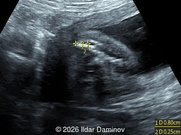
Our imaging demonstrates a fetal sacrococcygeal formation measuring 8.0 mm in length and 2.5 mm in width within the subcutaneous tissues. No associated intracranial or spinal anomalies were detected. A healthy male infant was delivered at 39 weeks via repeat C-section (3,720 g, 53 cm, Apgar 8-9). Postnatal clinical examination confirmed a benign, blind-ending sacral dimple with no deep or neural communication.
- Image 1 and Videos 1, 2 show a hyperechoic subcutaneous formation measuring 8 x 2.5 mm within the sacrococcygeal tissues at 21 weeks and 2 days of gestation. Sagittal and coronal views demonstrate intact spine anatomy with no associated osseous or structural vertebral anomalies.
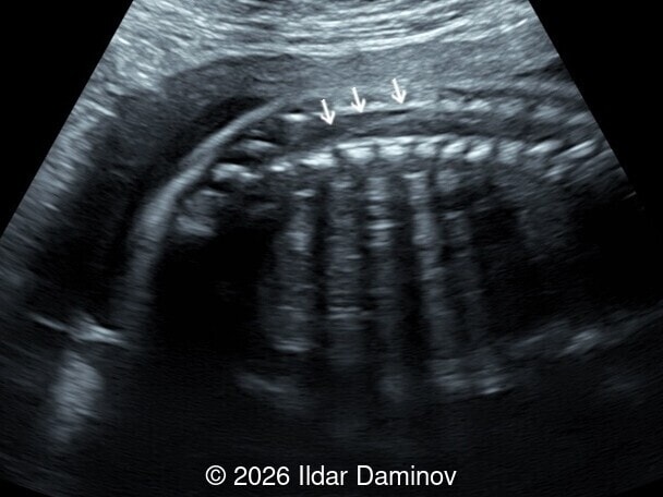
- Image 2: Sagittal view of the fetal spine at 25 weeks, demonstrating a normally positioned conus medullaris at the L1-L2 level, excluding tethered cord syndrome.
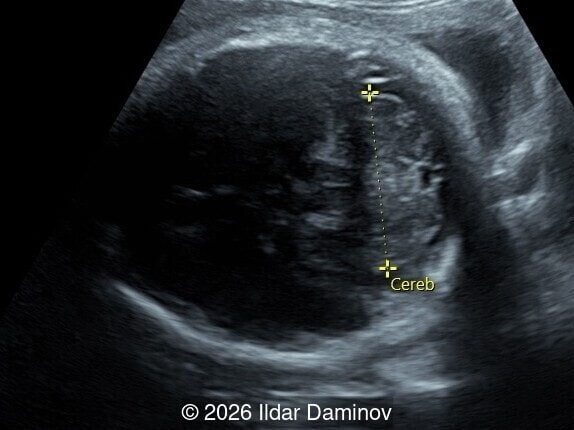
- Image 3: Axial view of the fetal head at 33 weeks, demonstrating a normal cerebellum and intact posterior fossa.
- Video 3: Axial view demonstrating a hyperechoic subcutaneous formation within the sacrococcygeal tissues and a normally developed fetal anal sphincter.
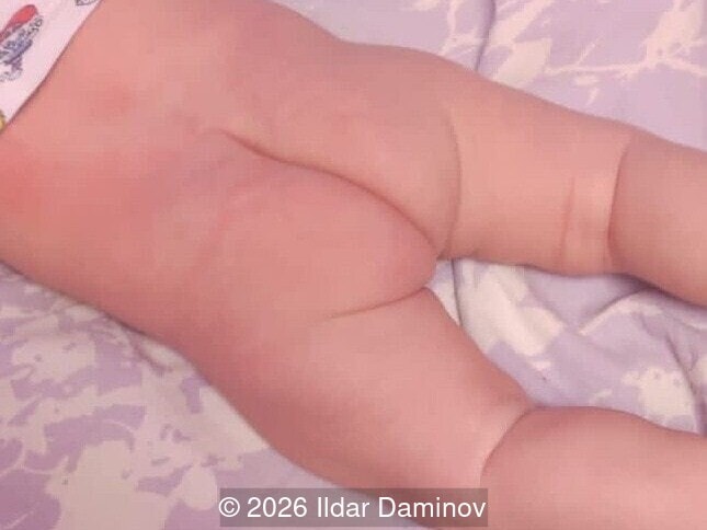
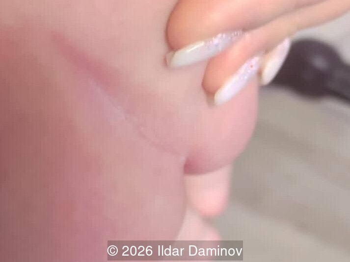
Postnatal clinical images of the neonate confirm a deep, isolated sacral dimple located within the upper intergluteal cleft. External visual inspection demonstrates completely intact overlying skin with a blind fundus and no signs of fistulous communication, discharge, or associated cutaneous stigmata.
Discussion
An isolated sacral dimple is a benign midline cutaneous invagination located within the intergluteal cleft, present in approximately 2% of healthy newborns [1]. The primary clinical significance of a sacrococcygeal finding lies in the necessity to differentiate these benign variants from markers of closed spinal dysraphism [2]. In a closed spinal dysraphism there is intact tissue covering the spinal defect although anomalies such as lipomas, fibrous bands or sinus tracts can lead to tethered cord syndrome. When left untreated, tethered cord syndrome follows a progressive course, with gradual worsening of neurologic and orthopedic functions [2]. Progressive traction on the spinal cord can lead to symptoms such as lower-extremity weakness or spasticity, gait disturbances, paresthesia, bowel or bladder dysfunction, back or leg pain, and development of scoliosis or foot abnormalities such as pes cavus [2,3]. Findings on MRI that can lead to tethered cord includes lipomyelomeningocele, conus lipoma, filar lipoma, tight filum terminale, split cord malformation and dermal sinus tract which is an epithelial lined channel extending to intrathecal structures and additionally can serve as a conduit for infection such as meningitis [2]. Embryologically, the caudal spinal cord develops through secondary neurulation, during which the caudal cell mass undergoes canalization and subsequent differentiation. Abnormalities of caudal neurulation and disjunction between the surface and neural ectoderm can result in occult spinal dysraphism and associated cutaneous markers [2].
A sacral dimple is classified as isolated and low-risk when it is small (<5 mm in diameter), located low in the intergluteal cleft (<2.5 cm from the anal verge), has a fully visible blind fundus, and lacks associated cutaneous markers such as hypertrichosis, lipomas, or vascular stains [2,3]. The reported incidence of abnormal underlying pathology in healthy neonates with isolated sacral dimple is 2%, though only 0.13% require surgical treatment for tethered cord syndrome [4]. High risk sacral dimples are large (>5mm), multiple, deep, >2.5cm from the anus, off midline, and associated with cutaneous stigmata such as localized hypertrichosis, hemangiomas, skin tags, lipomas, rudimentary tails, or pigmentary abnormalities [2,3]. In these cases, approximately 40% are associated with occult spinal anomalies [3]. Due to the low risk of occult spinal dysraphism in isolated sacral dimple, several authors argue that routine postnatal screening with ultrasound is not necessary [2,3].
There are no known reports of prenatal diagnosis of isolated sacral dimple. In our case, documentation of a normal conus medullaris position and unremarkable cranial markers in utero allowed for a confident diagnosis, providing parental reassurance and preventing unnecessary neonatal MRI or invasive testing. Postnatal external clinical evaluation of the male neonate confirmed a deep, blind-ending sacral dimple with completely intact skin and no communication with the spinal canal. This case emphasizes the clinical importance of correlating high-resolution prenatal ultrasound with postnatal clinical inspection to confidently confirm benign variants and reassure parents.
References
- Solak SS, Altunay IK, Demirci GT, et al. Prevalence of Congenital Cutaneous Anomalies in 1000 Newborns and a Review of the Literature. Am J Perinatol. 2016 Jan;33(1):79-83
- Eun J, Lee KS, Yang SH. Sacral dimple: clinical perspectives of lesions hidden beneath the skin. Clin Exp Pediatr. 2025 Nov 26;69(2):103–113
- Kriss VM, Desai NS. Occult spinal dysraphism in neonates: assessment of high-risk cutaneous stigmata on sonography. AJR Am J Roentgenol. 1998 Dec;171(6):1687-92.
- Kucera JN, Coley I, O'Hara S, et al. The simple sacral dimple: diagnostic yield of ultrasound in neonates. Pediatr Radiol. 2015 Feb;45(2):211-6.