Case of the Week # 659
(1) Department of Obstetrics and Gynecology (Head: Karol Dókuš, MD, PhD), F. D. Roosevelt University Hospital, Banská Bystrica, Slovakia; (2) Department of Medical Genetics, F. D. Roosevelt University Hospital, Banská Bystrica, Slovakia
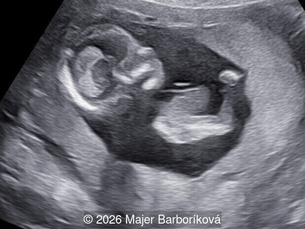
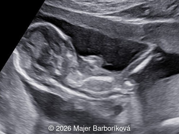
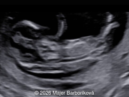
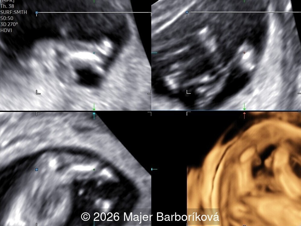
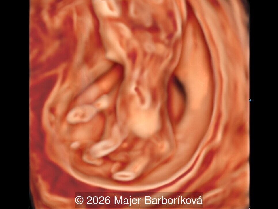
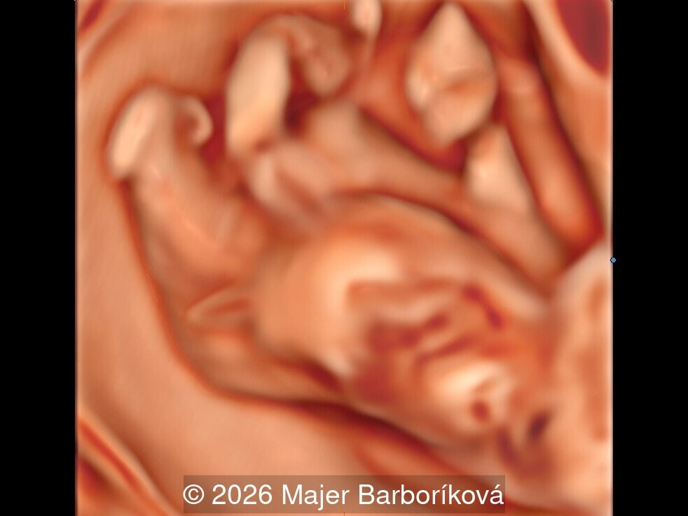
A 40-year-old primigravida with an unremarkable personal and family history was referred to our department at 12 weeks and 4 days of gestation because of an abnormal first-trimester scan. The pregnancy had been uncomplicated. The ultrasonographic evaluation revealed the following findings:
View the Answer Hide the Answer
Answer
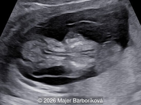
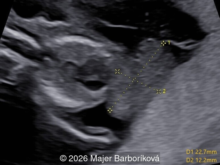
We present a case of Sirenomelia associated with a large omphalocele, also known as the mermaid syndrome.
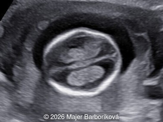
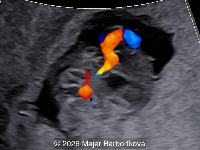
Transabdominal ultrasound examination at 12 weeks and 4 days of gestation, with a crown-rump length of 56 mm, demonstrated a large defect of the anterior abdominal wall with an omphalocele containing the liver, a single umbilical artery, and lower limbs fused into a single extremity with hypoplastic feet. The upper limbs appeared normal. The nuchal translucency measured 1.0 mm, the nasal bone was present and the intracranial translucency was normal, thus the markers of open spina bifida were negative. The patient was counseled about the diagnosis and the lethal prognosis, and referred for genetic consultation.
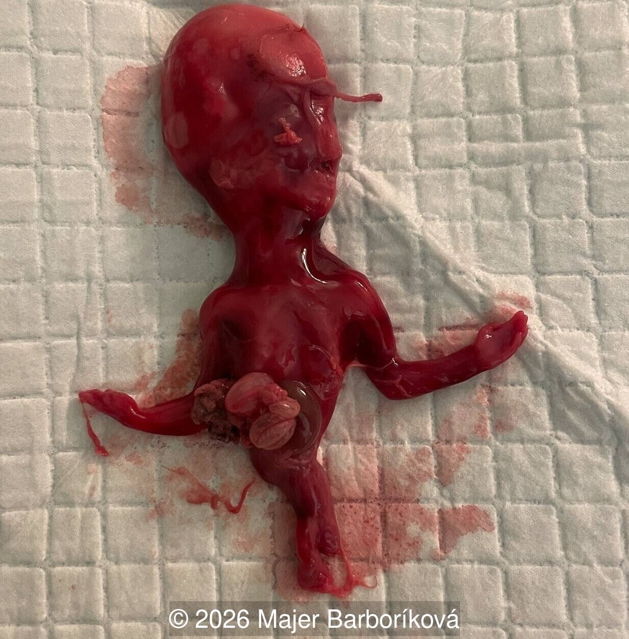
She returned at 15 weeks of gestation for amniocentesis. The scan performed at that time confirmed the previous findings: the urinary bladder was visualized, the kidneys could not be reliably assessed and renal dysgenesis was suspected in the context of the diagnosis, although it could not be confirmed by ultrasound. The amniotic fluid volume was still normal for gestational age. Amniocentesis was attempted but unsuccessful. After further counseling, the couple elected termination of pregnancy, which was performed at 15 weeks by prostaglandin induction and followed by curettage of the uterine cavity for a retained placenta. Photographic documentation of the fetus after termination confirmed the prenatal findings, with normally formed upper limbs, a large ventral wall defect with externalized abdominal contents, and lower limbs fused proximally into a single midline extremity with two hypoplastic feet distally. Genetic analysis was performed on fetal tissue. Quantitative fluorescent polymerase chain reaction showed a male fetus with a diploid complement of chromosomes 13, 15, 16, 18, 21, 22, X and Y, with no numerical chromosomal aberration, and Beckwith-Wiedemann syndrome was subsequently excluded.
Discussion
Sirenomelia is a rare and lethal malformation, with a prevalence of approximately 1 per 100,000 births [1]. It is usually attributed to the vitelline artery steal mechanism, in which an aberrant vessel arising high in the abdominal aorta diverts blood away from the caudal part of the embryo. The single umbilical artery is the sonographic evidence of that mechanism [2]. The classic picture of fused limbs, bilateral renal agenesis, an absent bladder and anhydramnios may be incomplete in early gestation. At 12 weeks, the kidneys are difficult to evaluate. The bladder is normally visible from about 11 weeks, and the amniotic fluid volume is not yet a useful marker, since renal oligohydramnios generally appears only after 16 weeks [3]. Renal involvement is also variable and ranges from agenesis to dysgenesis, so a visualized bladder does not exclude the diagnosis [1]. What can be assessed reliably is the contour of the lower body and the number of vessels in the umbilical cord, and these carried the diagnosis in our case. Normal markers do not exclude a severe malformation: in our patient both the nuchal and the intracranial translucency were normal. The intracranial translucency is a marker of open spina bifida, and sirenomelia is a disorder of the caudal blastema rather than an open neural tube defect, so the posterior fossa is expected to be normal [4]. The large omphalocele widens the differential diagnosis, in particular towards limb-body wall complex and the OEIS complex (Omphalocele, Exstrophy, Imperforate anus, Spinal defects), in which the bladder is not visualized and the lower limbs are separate. Early diagnosis is feasible in experienced hands [5,6], and the first trimester is the most favorable window, because later in gestation severe oligohydramnios obscures the fetus.
Sirenomelia is almost uniformly lethal. Most affected fetuses die in utero or shortly after birth from pulmonary hypoplasia and renal failure associated with bilateral renal agenesis. A small number of long-term survivors has been reported. In these cases, patients have at least one functioning kidney and a milder degree of caudal involvement which requires a series of reconstructive procedures to separate the limbs. Survival therefore depends on the degree of renal involvement and on the extent of the associated anomalies [1].
References
- Orioli IM, et al. Sirenomelia: an epidemiologic study in a large dataset from the International Clearinghouse of Birth Defects Surveillance and Research, and literature review. Am J Med Genet C Semin Med Genet. 2011;157C(4):358-373.
- Stevenson RE, et al. Vascular steal: the pathogenetic mechanism producing sirenomelia and associated defects of the viscera and soft tissues. Pediatrics. 1986;78(3):451-457.
- Sepulveda W, et al. Sirenomelia sequence versus renal agenesis: prenatal differentiation with power Doppler ultrasound. Ultrasound Obstet Gynecol. 1998;11(6):445-449.
- Chaoui R, et al. Assessment of intracranial translucency (IT) in the detection of spina bifida at the 11-13-week scan. Ultrasound Obstet Gynecol. 2009;34(3):249-252.
- Schiesser M, et al. Sirenomelia, the mermaid syndrome: detection in the first trimester. Prenat Diagn. 2003;23(6):493-495.
- Sepulveda W, et al. First-trimester sonographic diagnosis of sirenomelia: a multicenter series of 12 cases and review of the literature. Prenat Diagn. 2020;40(5):626-634.
Discussion Board
Winners

Alexandr Krasnov Ukraine Physician
Murat Cagan Turkey Physician

Ionut Valcea Romania Physician

Annette Reuss Germany Physician

Sruthi Pydi India Physician

Mert Eyupoglu Turkey Physician

Gulten Rafibeyli Azerbaijan Physician

Elnara Baghirova Azerbaijan Physician
Gianmarco Taraschi Canada Physician