Case of the Week # 658
MIRAS Medical Clinic, Sterlitamak, Republic of Bashkortostan, Russia
A 38-year-old G3P2 woman without significant medical history presented to our center for a routine third-trimester screening at 35 weeks of gestation.
View the Answer Hide the Answer
Answer
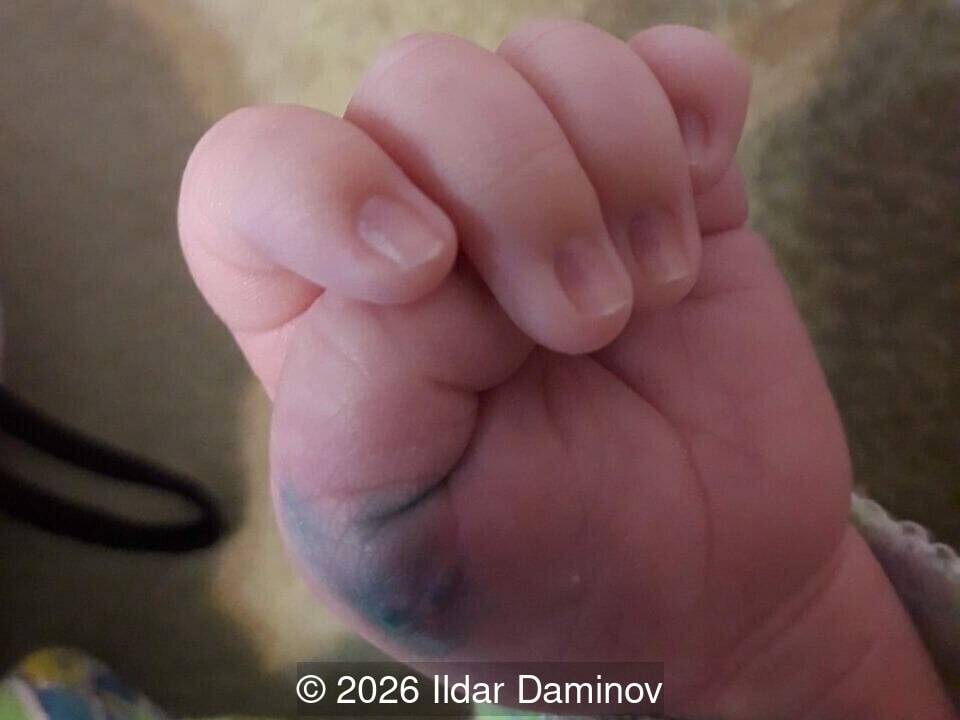
We present a case of bilateral preaxial polydactyly with intrauterine necrosis of the left accessory digit. Following birth, the child underwent successful two-stage surgical treatment.
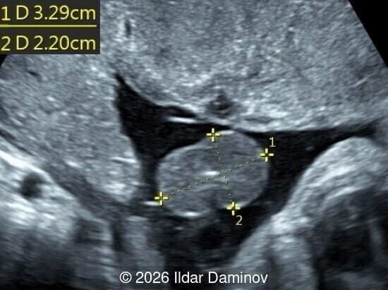
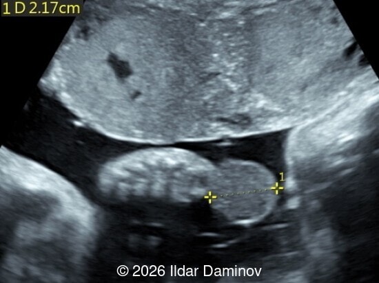
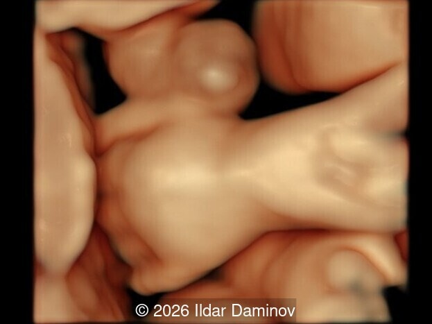
- Images 1-3 and Video 1 demonstrate a tumor-like mass on the left hand with its sonographic measurements, structurally represented by the intrauterine necrosis of an extra digit. The lesion presents at the base of the duplicated thumb as a solid, well-circumscribed, and rounded soft-tissue mass measuring 33 × 22 × 22 mm. It exhibits homogeneous internal echogenicity without evidence of calcification, consistent with an edematous rudimentary digit undergoing antenatal tissue remodeling.
- Video 2 shows minimal central blood flow within the mass on Color Doppler, consistent with subtotal ischemic remodeling during intrauterine necrosis of the extra digit.
- Video 3, in slow motion, demonstrates the presence of a right-hand preaxial polydactyly without structural alterations of the digit.
Discussion
Polydactyly is a common congenital limb anomaly characterized by the presence of extra digits on the hands or feet, which can range from complex osseous structures to simple soft-tissue tags attached by a narrow pedicle. Polydactyly occurs with a prevalence of approximately 23 per 10,000 live births [1]. It can be classified by position of the accessory digit: (1) preaxial: located on the radial side of the hand with duplication of the thumb, (2) central: duplication of the index, middle, or ring finger, and (3) postaxial: located on the ulnar side of the hand with duplication of the small finger [2]. In type A the extra digit is well developed, whereas in type B the extra digit is rudimentary, without skeletal structure [3]. Polydactyly is associated with other anomalies in approximately 15% of cases [4]. Of the polydactyly cases with an identified syndrome, Meckel-Gruber, Trisomy 13 and 21 are the most common [4]. Therefore, in patients with polydactyly, a detailed ultrasonographic examination and fetal karyotyping should be performed to evaluate for associated anomalies and syndromes [3]. Isolated postaxial polydactyly often follows an autosomal dominant inheritance pattern [5].
When dealing with a rudimentary soft-tissue digit, the thin neurovascular bundle is highly susceptible to spontaneous torsion or constriction in utero, and could be triggered by fetal movements [2,6] including sucking the accessory digit [7] or amniotic bands [8]. If this occurs during gestation, the interruption of arterial blood supply leads to prenatal ischemic necrosis of the accessory digit. Depending on the timing of the vascular accident before delivery, the fetus may be born with a gangrenous mass at the site of the accessory digit [6,9] or, with a remnant bump on the side of the hand due to spontaneous amputation [2,3]. The auto-amputated digit can be visualized in the amniotic cavity by ultrasound [2]. In a study by Zimmer et al describing 16 fetuses with isolated polydactyly, 9 were born with a small bump on the hand suggesting that in utero partial autoamputation occurs commonly [3]. Two of these cases had bilateral polydactyly in which one hand had a small bump and the other hand was normal, suggesting complete, spontaneous amputation of the accessory digit [3].
Prenatal detection of spontaneous loss of an accessory digit is rare with only two prenatal cases described in the literature [2,10]. In one case, unilateral type A postaxial polydactyly was detected at 13 weeks gestation and autoamputation demonstrated at 19 weeks [10]. In the second case, unilateral type B postaxial polydactyly was identified at 18 weeks and autoamputation at 20 weeks gestation [2]. For prenatal ultrasound specialists, the detection of a changing, solid, tumor-like mass on a fetal hand requires a meticulous evaluation to distinguish from other conditions such as congenital hemangioma [11], myofibroma [12], infantile fibrosarcoma [13], rhabdomyosarcoma [14], and other rare soft-tissue anomalies. A detailed targeted evaluation of the contralateral limb is crucial and the discovery of a healthy accessory digit on the opposite hand suggests that the tumor-like mass is a necrotic extra digit, rather than a prenatal tumor. Accurate prenatal diagnosis of intrauterine polydactyly complicated by vascular torsion and necrosis is valuable for patient counselling.
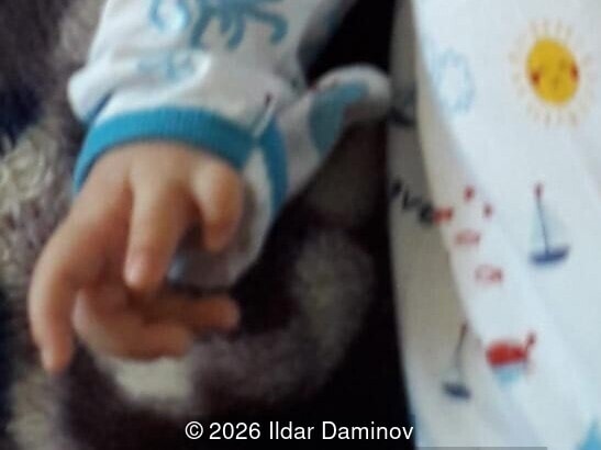
In the present case, the patient required a two-stage surgical management due to the bilateral nature of the anomaly. The first stage was performed immediately after delivery to excise the tumor-like necrotic mass on the affected left hand. Because this intrauterine vascular accident naturally thromboses the feeding vessels, the devitalized tissue can be excised after delivery with minimal trauma and risk of bleeding [9]. The second stage involved the elective surgical removal of the healthy accessory digit on the contralateral hand, which was performed at a later stage.
References
- Goldfarb CA, Shaw N, Steffen JA, et al. The Prevalence of Congenital Hand and Upper Extremity Anomalies Based Upon the New York Congenital Malformations Registry. J Pediatr Orthop. 2017 Mar;37(2):144-148.
- Volpe N, Franchi L, Mazzone E, et al. Self-Amputation of the Extra Digit in a Fetus with Polydactyly: First Ultrasound Demonstration. Fetal Diagn Ther. 2017;41(4):314-316.
- Zimmer EZ, Bronshtein M. Fetal polydactyly diagnosis during early pregnancy: clinical applications. Am J Obstet Gynecol. 2000 Sep;183(3):755-8.
- Castilla EE, Lugarinho R, da Graça Dutra M, et al. Associated anomalies in individuals with polydactyly. Am J Med Genet. 1998 Dec 28;80(5):459-65.
- Galjaard RJH, Smits APT, Tuerlings JHAM, et al. A new locus for postaxial polydactyly type A/B on chromosome 7q21-q34. Eur J Hum Genet. 2003 May;11(5):409-15.
- Watanabea D, Hasebea Y, Mitsuib H, et al. Prenatal Torsion of Radial Polydactyly: A Gangrenous Mass at the Base of the Thumb. Acta Med Okayama. 2023 Dec;77(6):651-653.
- Kanter WR, Upton J. "Pacifier polydactyly": a transitional form between pedunculated polydactyly and rudimentary polydactyly. Plast Reconstr Surg. 1989 Jul;84(1):136-9.
- Chung P. EP11.18: Prenatal diagnosis of amniotic band syndrome: a case report. Ultrasound Obstet Gynecol. 2018 Oct; 52: 248-248.
- Saraf S. Intra-natal Torsion of Polydactyly. J Cutan Aesthet Surg. 2011 Jan;4(1):56-7.
- Porozova E. EP06.08: A rare case of prenatal auto-amputation of extra-finger in fetus with isolated postaxial polydactyly diagnosed at 13 weeks of pregnancy. Ultrasound Obstet Gynecol. 2015;46(S1):203-204.
- Delgadillo D, Carr LW, Brgoch MS, et al. Rapidly Involuting Congenital Hemangioma of the Hand. Eplasty. 2017 Mar 8;17:ic6.
- Melikoglu C, Keklik B, Sutcu M, et al. Infantile myofibroma: A differential diagnosis of hand tumors during the neonatal period. J Pediatr Surg Case Rep. 2014 May;2(5):222-224.
- Kimura C, Kitamura T, Sugihara T. A case of congenital infantile fibrosarcoma of the right hand. J Dermatol. 1998 Nov;25(11):735-41.
- Khallouki M, Aboudourib M, Bendaoud L, et al. Spindle Cell Rhabdomyosarcoma of the Hand in a Full-Term Newborn: A Case Report. Cureus. 2025 Feb 26;17(2):e79670.
Discussion Board
Winners

Elena Andreeva Russian Federation Physician

YULIA VISHNEVSKAYA Russian Federation Physician

Gulten Rafibeyli Azerbaijan Physician