Case of the Week # 657
(1) Department of Genetics, Polish Mother's Memorial Hospital, Lodz, Poland; (2) Laboratory of Cytogenetics, Department of Medical Genetics, Institute of Mother and Child, Warsaw, Poland; (3) Centro Médico Recoletas, Valladolid, Spain
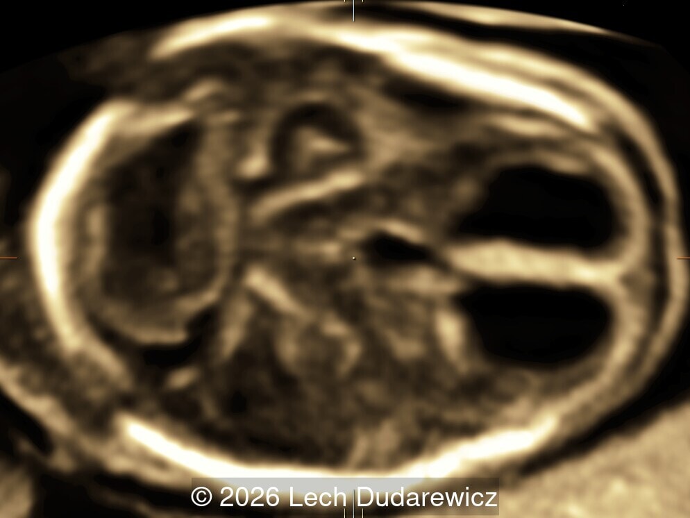
This fetus was examined at the end of the first trimester because of markedly increased nuchal translucency. The crown-rump length corresponded to approximately 13 weeks of gestation, and the nuchal translucency measured 7.5 mm. Targeted early neurosonography was performed.
View the Answer Hide the Answer
Answer
We present a case of thanatophoric dysplasia type II, caused by the pathogenic FGFR3 K650E variant. The diagnosis was confirmed by exome sequencing performed on trophoblast DNA, which identified a pathogenic heterozygous variant in FGFR3 (NM_000142.5:c.1948A>G; p.Lys650Glu / K650E).
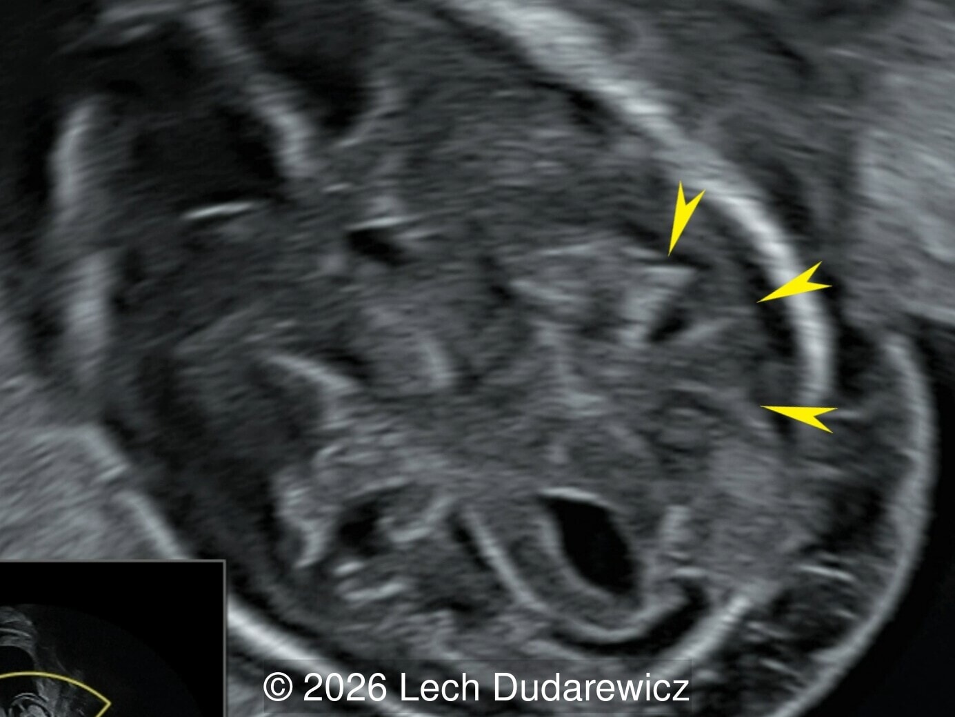
Our ultrasound images demonstrated the following findings: Video 1 shows short, mildly bent long bones and a narrow thorax caused by short ribs — in particular shortening of the ossified rib portions — together with relative cardiomegaly, macrocrania, increased nuchal translucency, and lateral neck cysts. Video 2 shows the fetal brain in a low axial/transverse plane at the end of the first trimester; the yellow arrows (Image 1) indicate abnormal radial echogenic sulci in the temporal regions, consistent with temporal lobe dysplasia. Because temporal sulcation should not yet be visible at this gestational age, this represents abnormal, premature sulcation. Brainstem hypoplasia with a wide cerebral aqueduct due to the abnormal brainstem configuration, was also present. The molecular result — the FGFR3 K650E variant, strongly associated with type II — confirms the diagnosis and explains the combined skeletal, cranial, and cerebral phenotype [1]. Despite the presence of FGFR3 mutation, which is strongly associated with thanatophoric dysplasia type II, there was no kleeblattschädel deformity, and the long bones were only mildly bowed. This may simply reflect the very early gestational age at presentation.
Discussion
Thanatophoric dysplasia is the most common lethal skeletal dysplasia and results from de novo activating mutations in FGFR3 [1]. Two types of thanatophoric dysplasia, types 1 and 2, are distinguished based on the shape of the skull and the morphology of the femur. Thanatophoric dysplasia type 1 is characterized by micromelia with bowed femurs and, less commonly, craniosynostosis of variable severity. Type 2 is classically characterized by marked micromelia, a narrow thorax, relatively straight (rather than curved) long bones, and a cloverleaf skull due to severe craniosynostosis, and is typically caused by the FGFR3 p.Lys650Glu (K650E) variant identified in this fetus [1]. This case illustrates that the brain phenotype can provide an important diagnostic clue very early in pregnancy.
An important finding that leads directly to the diagnosis of this skeletal dysplasia is the very early‑onset migration anomaly of the temporal lobe, which begins at least as early as 13.5 weeks of gestational age [2]. Shortly after thanatophoric dysplasia was described as an independent entity by Maroteaux et al. [3] in 1967, Goutières, Aicardi, and Farkas‑Bargeton [4] reported in 1971 a cerebral malformation affecting the temporal cortex in two term infants with thanatophoric dwarfism. Since then, various brain anomalies have been documented in postmortem examinations ranging in age from the 15th week of gestation to term, with temporal lobe dysplasia being one of the most consistently observed brain abnormalities, both prenatally and postnatally, in thanatophoric dysplasia [2, 5-15]. This dysplasia is characterized by hippocampal dysplasia (100% of cases), temporal lobe enlargement (93%), and deep transverse sulci with broad gyri oriented transversely across the medial and inferior temporal surfaces (88%). Other features include rudimentary dentate gyrus (100%), polymicrogyria (97%), subependymal and/or subarachnoid neuronal heterotopia (81% and 79%, respectively), hydrocephalus (56%), cerebellar cortex abnormalities (37%), and hypoplasia or partial agenesis of the corpus callosum (34%) [11]. Vogt and Blaas [13] described the necropsy findings of 25 cases of thanatophoric dysplasia, identifying temporal lobe dysplasia in 52% of the entire series and in 100% of the cases examined from 1998 onwards. The authors attribute this temporal increase primarily to the lack of earlier awareness of the condition. In this regard, Ralph Waldo Emerson’s well‑known assertion—familiar to followers of Philippe and TheFetus.net—aptly applies: “People only see what they are prepared to see.” There are no noticeable differences in central nervous system abnormalities between thanatophoric patients with and without cloverleaf skull [7].
FGFR3 plays a key role in bone development, where activation of the RAS/ERK pathway exerts an inhibitory effect on chondrocyte proliferation in the growth plate cartilage, resulting in the severe impairment of bone ossification observed in thanatophoric dysplasia [16]. Except for hypoplasia of the posterior fossa and narrowing of the foramen magnum with occasional brainstem compression and hydrocephalus, there is no morphological evidence that the central nervous system abnormalities are caused by skull defects (platybasia and craniosynostosis) [11]. FGFR3 mutations activate the receptor tyrosine kinase [17], which also disrupts processes involved in cerebral cortical development. The malformation in thanatophoric dysplasia arises from a combination of alterations in cortical patterning, proliferation, and apoptosis affecting the cerebral cortex. The cortical surface undergoes marked expansion at the onset of development of the future temporal lobe, beginning between 6 and 8 weeks of gestation. This early expansion initially leads to enlargement of the lateral ventricle and subsequently causes the cortex to fold, forming aberrant deep transverse sulci. Furthermore, at the beginning of cortical development, the hippocampus exhibits an abnormal pattern, possibly due to a combination of suppressed activity of the cortical hem (an embryonic structure located along the medial border of the dorsal telencephalon) and direct effects of constitutive FGFR3 activation [11].
Although central nervous system abnormalities have been widely recognized in postmortem examinations, they have not been frequently described in fetal imaging. However, multiple deep fissures along the inferomedial temporal and occipital surfaces, as well as megalencephaly, can be identified using ultrasound or magnetic resonance imaging [18,19]. Similar cortical‑level findings have been reported in other skeletal dysplasias caused by mutations in the FGFR3 gene (achondroplasia, both pre‑ and postnatally, and hypochondroplasia postnatally) [20-23]. In thanatophoric dysplasia, abnormal sulcation typically involves the inferior aspect of the temporal lobes, whereas in achondroplasia and hypochondroplasia the abnormal sulcation primarily affects the medial aspect of the temporal and occipital lobes [20]. In addition, expansion and over‑convolution of the temporal lobes, as well as temporal lobe clefts, have been described both pre‑ and postnatally in patients with Apert and Pfeiffer syndromes, findings that are particularly evident during the developmental period when the normal brain is still relatively smooth (approximately between 24 and 28 weeks of gestation) [24,25]. Both syndromes (Apert and Pfeiffer) are craniosynostoses caused by mutations in the FGFR2 gene, underscoring the role of FGF activation not only in osteogenesis but also in central nervous system development.
The key finding in this case is abnormal sulcation of the temporal regions. Temporal lobe dysplasia is a characteristic brain malformation of thanatophoric dysplasia and, on ultrasound, appears as abnormal prominent linear echogenic sulci along the medial or inferomedial temporal and occipital lobes [18,19,26,27]. These features are strikingly different from those of the normal 18‑ to 22‑week brain, whose surface is essentially smooth. The present case demonstrates that the same abnormality may already be visible at the end of the first trimester, when temporal sulcation is even less apparent. In the cohort reported by Wang et al. [27], temporal lobe dysplasia was initially identified in only 25% of prenatal ultrasound scans, a finding likely related both to lack of awareness of this feature (all diagnosed cases occurred after 2007, when the abnormality was first described) and to the fact that standard prenatal imaging planes do not allow optimal visualization of the temporal lobes. To improve diagnostic capabilities, these authors propose obtaining two additional planes that enhance visualization of the temporal lobe: a low axial plane below the level of the BPD plane, roughly including the orbits and brainstem/midbrain, and a coronal plane that also includes the cerebellum or midbrain. In addition, three‑dimensional imaging with multiplanar reconstructions can assist in achieving these specific views. Blaas et al. [18] further indicate that visualization of temporal lobe dysplasia can also be achieved using a parasagittal view that examines the medial periventricular cortex.
In addition to the brain abnormalities previously described in imaging studies, thanatophoric dysplasia has been reported in association with other central nervous system pathologies, including partial agenesis of the corpus callosum, encephalocele, semilobar holoprosencephaly, hydrancephaly, and enlargement of the cerebellum and cisterna magna [28-31]. In this fetus, brainstem hypoplasia and a wide aqueduct were also present. These findings are consistent with those reported by Tonni et al. [32], who described increased nuchal translucency, dysmorphic choroid plexuses, and early hydrocephalus as first‑trimester ultrasound markers of de novo thanatophoric dysplasia type II with cloverleaf skull.
The main differential diagnosis includes other lethal skeletal dysplasias that present with micromelia and a narrow thorax, such as severe achondroplasia, achondrogenesis, and osteogenesis imperfecta type II [33]. Differentiating thanatophoric dysplasia from achondroplasia can be challenging, as achondroplasia may exhibit similar brain abnormalities; however, the degree of micromelia is typically much more pronounced in thanatophoric dysplasia. Achondrogenesis and osteogenesis imperfecta type II can also manifest with severe limb shortening and a narrow thorax, but neither condition accounts for the characteristic temporal lobe dysplasia, and osteogenesis imperfecta additionally shows fractures and poor mineralization. Short‑rib thoracic dysplasias (skeletal ciliopathies) may produce a narrow thorax, short limbs, and brain findings.
In conclusion, abnormal temporal lobe sulcation is a characteristic neuroimaging feature of thanatophoric dysplasia and should be actively evaluated during fetal assessment when severe skeletal dysplasia is suspected. The presence of visible temporal sulcation at the end of the first trimester is abnormal and may represent an early marker of temporal lobe dysplasia. Recognition of these intracranial abnormalities can complement the skeletal phenotype, improving diagnostic confidence and supporting earlier prenatal diagnosis of thanatophoric dysplasia.
References
- Thanatophoric Dysplasia. In: GeneReviews®. Seattle (WA): University of Washington, Seattle; 1993–2024. Available from: https://www.ncbi.nlm.nih.gov/books/NBK1366/
- Knisely AS, Ambler MW. Temporal-lobe abnormalities in thanatophoric dysplasia. Pediatr Neurosci. 1988;14(4):169-176.
- Maroteaux P, Lamy M, Robert JM. Le nanisme thanatophore. Presse Med (1893). 1967 Nov 22;75(49):2519-2524.
- Goutieres F, Aicardi J, Farkas-Bargeton E. Une malformation cérébrale particulière associée au nanisme thanatophore. Presse Med (1893). 1971 Apr 24;79(21):960.
- Wongmongkolrit T, Bush M, Roessmann U. Neuropathological findings in thanatophoric dysplasia. Arch Pathol Lab Med. 1983 Mar;107(3):132-135.
- Hori A, Friede RL, Fischer G. Ventricular diverticula with localized dysgenesis of the temporal lobe in cloverleaf skull anomaly. Acta Neuropathol. 1983;60(1-2):132-136.
- Ho KL, Chang CH, Yang SS, Chason JL. Neuropathologic findings in thanatophoric dysplasia. Acta Neuropathol. 1984;63(3):218-228.
- Shigematsu H, Takashima S, Otani K, Ieshima A. Neuropathological and Golgi study on a case of thanatophotoric dysplasia. Brain Dev. 1985;7(6):628-632.
- Coulter CL, Leech RW, Brumback RA, Schaefer GB. Cerebral abnormalities in thanatophoric dysplasia. Childs Nerv Syst. 1991 Feb;7(1):21-26.
- Yamaguchi K, Honma K. Autopsy case of thanatophoric dysplasia: observations on the serial sections of the brain. Neuropathology. 2001 Sep;21(3):222-228.
- Hevner RF. The cerebral cortex malformation in thanatophoric dysplasia: neuropathology and pathogenesis. Acta Neuropathol. 2005 Sep;110(3):208-221.
- Itoh K, Pooh R, Kanemura Y, et al. Brain malformation with loss of normal FGFR3 expression in thanatophoric dysplasia type I. Neuropathology. 2013 Dec;33(6):663-666.
- Vogt C, Blaas HG. Thanatophoric dysplasia: autopsy findings over a 25-year period. Pediatr Dev Pathol. 2013 May-Jun;16(3):160-167.
- Shinde RR, Srinivasan L, Raja V, Seshadri S. Thanatophoric Dysplasia and the Brain—A Perinatal Pathology Study. J Fetal Med. 2018;5:145–149.
- Mondal A, Mandal B, Das RN, et al. Thanatophoric dysplasia type 1 with temporal lobe dysplasia: Report of a case along with differential diagnosis. Indian J Pathol Microbiol. 2021 Oct-Dec;64(4):776-779.
- Krejci P. The paradox of FGFR3 signaling in skeletal dysplasia: why chondrocytes growth arrest while other cells over proliferate. Mutat Res Rev Mutat Res. 2014 Jan-Mar;759:40-48.
- Naski MC, Wang Q, Xu J, Ornitz DM. Graded activation of fibroblast growth factor receptor 3 by mutations causing achondroplasia and thanatophoric dysplasia. Nat Genet. 1996 Jun;13(2):233-237.
- Blaas HG, Vogt C, Eik-Nes SH. Abnormal gyration of the temporal lobe and megalencephaly are typical features of thanatophoric dysplasia and can be visualized prenatally by ultrasound. Ultrasound Obstet Gynecol. 2012 Aug;40(2):230-234.
- Fink AM, Hingston T, Sampson A, et al. Malformation of the fetal brain in thanatophoric dysplasia: US and MRI findings. Pediatr Radiol. 2010 Dec;40 Suppl 1:S134-S137.
- Pugash D, Lehman AM, Langlois S. Prenatal ultrasound and MRI findings of temporal and occipital lobe dysplasia in a twin with achondroplasia. Ultrasound Obstet Gynecol. 2014 Sep;44(3):365-368.
- Manikkam SA, Chetcuti K, Howell KB, et al. Temporal Lobe Malformations in Achondroplasia: Expanding the Brain Imaging Phenotype Associated with FGFR3-Related Skeletal Dysplasias. AJNR Am J Neuroradiol. 2018 Feb;39(2):380-384.
- Linnankivi T, Mäkitie O, Valanne L, Toiviainen-Salo S. Neuroimaging and neurological findings in patients with hypochondroplasia and FGFR3 N540K mutation. Am J Med Genet A. 2012 Dec;158A(12):3119-3125.
- Philpott CM, Widjaja E, Raybaud C, et al. Temporal and occipital lobe features in children with hypochondroplasia/FGFR3 gene mutation. Pediatr Radiol. 2013 Sep;43(9):1190-1195.
- Stark Z, McGillivray G, Sampson A, et al. Apert syndrome: temporal lobe abnormalities on fetal brain imaging. Prenat Diagn. 2015 Feb;35(2):179-182.
- Khonsari RH, Delezoide AL, Kang W, et al. Central nervous system malformations and deformations in FGFR2-related craniosynostosis. Am J Med Genet A. 2012 Nov;158A(11):2797-2806.
- Miller E, Blaser S, Shannon P, Widjaja E. Brain and bone abnormalities of thanatophoric dwarfism. AJR Am J Roentgenol. 2009 Jan;192(1):48-51.
- Wang DC, Shannon P, Toi A, et al. Temporal lobe dysplasia: a characteristic sonographic finding in thanatophoric dysplasia. Ultrasound Obstet Gynecol. 2014 Nov;44(5):588-594.
- Kalache KD, Lehmann K, Chaoui R, et al. Prenatal diagnosis of partial agenesis of the corpus callosum in a fetus with thanatophoric dysplasia type 2. Prenat Diagn. 2002 May;22(5):404-407.
- Li D, Liao C, Ma X, et al. Thanatophoric dysplasia type 2 with encephalocele during the second trimester. Am J Med Genet A. 2006 Jul 1;140(13):1476-1477.
- Martínez-Frías ML, Egüés X, Puras A, et al. Thanatophoric dysplasia type II with encephalocele and semilobar holoprosencephaly: Insights into its pathogenesis. Am J Med Genet A. 2011 Jan;155A(1):197-202.
- Chen CP, Chang TY, Lin TW, et al. Prenatal diagnosis of hydrancephaly and enlarged cerebellum and cisterna magna in a fetus with thanatophoric dysplasia type II and a review of prenatal diagnosis of brain anomalies associated with thanatophoric dysplasia. Taiwan J Obstet Gynecol. 2018 Feb;57(1):119-122.
- Tonni G, Palmisano M, Ginocchi V, et al. Dysmorphic choroid plexuses and hydrocephalus associated with increased nuchal translucency: early ultrasound markers of de novo thanatophoric dysplasia type II with cloverleaf skull (Kleeblattschaedel). Congenit Anom (Kyoto). 2014 Nov;54(4):228-232.
- Soares MI, Veríssimo P, Coelho C, et al. Thanatophoric Dysplasia Type I Confirmed by Fibroblast Growth Factor Receptor 3 (FGFR3) Mutation: Clinical Course and Ethical Considerations. Cureus. 2026 Apr 3;18(4):e106363.
Discussion Board
Winners

Andrii Averianov Ukraine Physician

Nguyen Thac Viet Viet Nam Physician

Ionut Valcea Romania Physician

Anette Beverdam Netherlands Sonographer

CHERYL TURNER United States Sonographer

Petra Majer Barboríková Slovakia Physician

Tetiana Ishchenko Ukraine Physician

Asmaa Rehan Egypt Physician
Noelia Bardón Spain Physician