Case of the Week # 656
(1) Rochester, NY, USA; (2) Centro Médico Recoletas, Valladolid, Spain
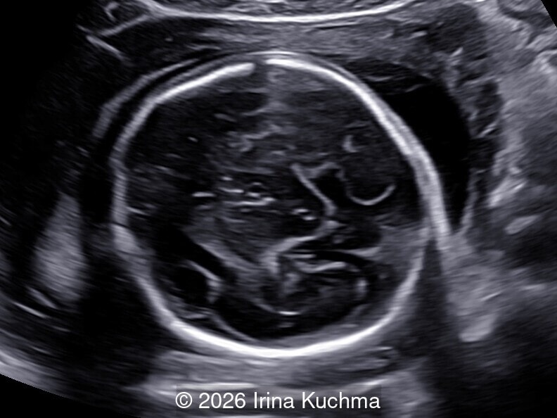
34-year-old woman G1P0 with no significant previous medical history presented for her 20 weeks ultrasound. Genetic test results were normal.
View the Answer Hide the Answer
Answer
We present a case of thanatophoric dysplasia. Patient opted to terminate the pregnancy.
Our ultrasound images demonstrate:
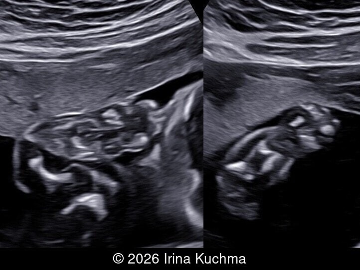
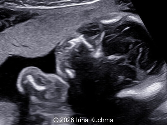
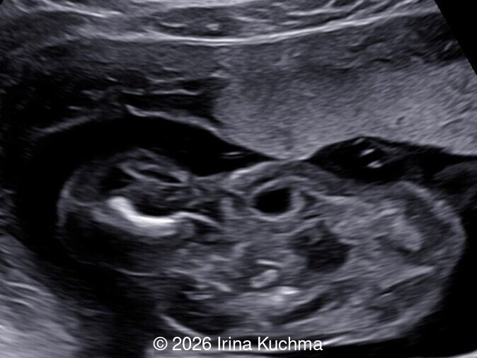
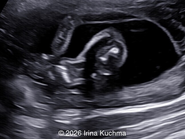
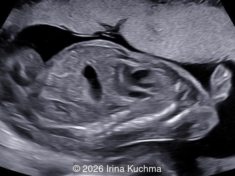
- Images 1-4: depict the humerus, radius and ulna, femur, tibia, and fibula, demonstrating marked shortening and bowing of the long bones.
- Image 5: sagittal image showing the narrow thorax compared to the normal-sized abdomen.
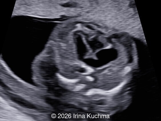
- Image 6: axial image of the chest showing a narrow thorax with the heart occupying most of the thoracic cavity.
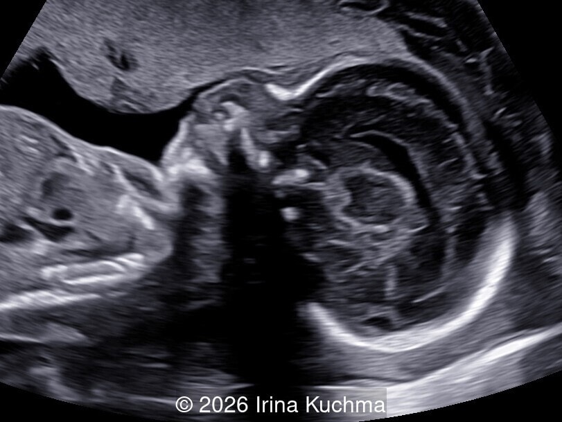
- Image 7: midsagittal view of the fetal head showing a large head and the beginning of frontal bossing.
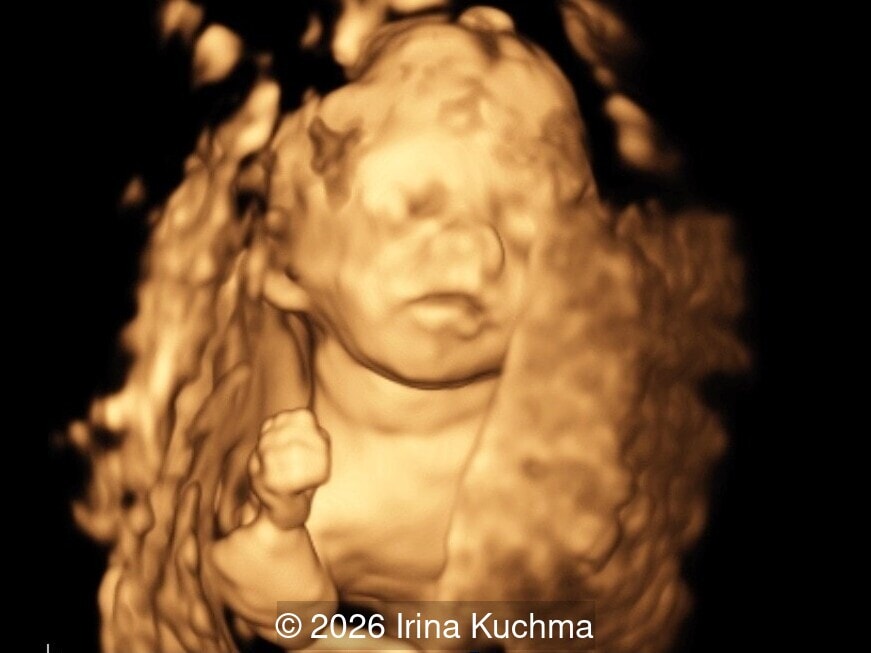
- Image 8: 3D rendering illustrating the characteristic facial dysmorphism and deformity of the right upper extremity.
- Image 9: axial view of the fetal head demonstrating relative macrocephaly.
Discussion
Fibroblast growth factor receptor 3 (FGFR3) disorders comprise a spectrum of pathologies within Group 1 of the 2023 revision of the nosology of genetic skeletal disorders. These include FGFR3 chondrodysplasias, such as thanatophoric dysplasia types 1 and 2, SADDAN (severe achondroplasia with developmental delay and acanthosis nigricans), achondroplasia, and hypochondroplasia, as well as syndromes with craniosynostosis, such as Crouzon syndrome with acanthosis nigricans and Muenke syndrome. Thanatophoric dysplasia is lethal, achondroplasia is non-lethal with rare exceptions, and hypochondroplasia may phenotypically resemble a mild form of achondroplasia [1].
The term thanatophoric dysplasia is derived from the Greek word thanatophoros, meaning “bearing death” [2]. Maroteaux et al. first described thanatophoric dwarfism in 1967, distinguishing it from achondroplasia [3]. Ten years later, the term thanatophoric dwarfism was replaced by thanatophoric dysplasia at the Second International Conference on the Nomenclature of Skeletal Dysplasias. Thanatophoric dysplasia is the most common lethal skeletal dysplasia and is caused by mutations in the FGFR3 gene, located on chromosome 4p16.3 [4]. The condition is inherited in an autosomal dominant manner, with reported birth prevalence ranging from 1:20,000 to about 1:50,000 [5,6]. The risk of FGFR3‑related disorders increases with advanced paternal age, as most cases result from de novo mutations arising in the paternally derived allele. This is attributed to the clonal expansion of PAE (paternal age effect) mutations in the testis, leading to a higher proportion of mutant sperm in older individuals [6,7].
There are two types of thanatophoric dysplasia based on two diagnostic criteria: the presence or absence of a cloverleaf skull, and the identification of either bowed or straight femurs. Thanatophoric dysplasia type 1 is characterized by micromelia with bowed femurs and, less commonly, craniosynostosis of variable severity. Thanatophoric dysplasia type 2 is characterized by micromelia with straight femurs and the consistent presence of moderate‑to‑severe craniosynostosis with a cloverleaf skull deformity [8]. A single recurrent mutation in the FGFR3 gene has been described for type 2 (p.Lys650Glu), whereas as many as 12 distinct missense mutations and several stop‑codon mutations have been identified in type 1, with p.Arg248Cys and p.Tyr373Cys accounting for approximately 90% of cases [9,10].
Thanatophoric dysplasia should be suspected in a fetus presenting with shortening of the long bones and increased nuchal translucency in the first trimester [11-13]. In the second or third trimester, the following findings can be seen: very short and often bowed long bones with good mineralization (telephone‑receiver appearance in type 1); well‑ossified spine and skull; platyspondyly (H‑shaped configuration on frontal view); narrow thorax with short ribs and a protuberant abdomen (“bell‑shaped” thoracoabdominal contour or “champagne cork” appearance); brachydactyly with trident configuration; relative macrocephaly with facial dysmorphism (flat nasal bridge and frontal bossing); and cloverleaf skull (more common in thanatophoric dysplasia type 2 than type 1) [1, 2, 14-17]. Polyhydramnios, often severe, is common in the third trimester. Additionally, a key finding described in FGFR3 chondrodysplasias is the early‑onset migration anomaly of the temporal lobe, visible as enlargement, abnormal gyration and sulcation, and polymicrogyria, accompanied by ventriculomegaly in more than 30% of cases [18]. According to the publication by Chitty et al., the overall frequency of the different findings is as follows: short femur (100%), narrow thorax (96%), frontal bossing (84%), increased nuchal translucency (80%), bowed femur (70%), macrocephaly (61%), increased amniotic fluid (59%), short fingers/trident hand (52%), and cloverleaf skull (30%) [17]. In the series reported by Rahemtullah et al., all cases of thanatophoric dysplasia exhibited a femur length‑to‑abdominal circumference ratio < 0.16, which was associated with a lethal outcome [19].
The diagnosis of thanatophoric dysplasia is highly reliable with 2D ultrasound, although 3D/4D ultrasound may add an additional dimension to patient counselling. The surface‑rendering mode facilitates direct visualization of fetal features, while the transparency/x‑ray modes are particularly useful for assessing the long bones and for visualizing the vertebrae and intervertebral spaces [15,20,21].
The first prenatal diagnosis of thanatophoric dysplasia using ultrasound was reported by Cremin and Shaff in 1977 [22]; although the condition had been identified a few years earlier on the basis of a plain abdominal radiograph [23,24]. The accuracy of prenatal diagnosis based on ultrasound findings has been reported to range from 40% to 88%, with detection rates exceeding 80% in the most recent series [17,25]. Confirmation of the diagnosis relies on molecular testing—targeted analysis, single‑gene testing, skeletal dysplasia multigene panels, or exome sequencing—performed through chorionic villus sampling, amniocentesis, or, more recently, analysis of cell‑free fetal DNA in maternal blood (with next‑generation sequencing outperforming PCR‑based methods) [2,4,17,26,27]. Establishing a definitive diagnosis is essential for accurate genetic counselling, enabling informed parental decision‑making regarding pregnancy management and future reproductive risk. The empiric recurrence risk in subsequent pregnancies is low, although slightly higher than that of the general population due to the possibility of parental mosaicism [28].
Potential pregnancy complications include prematurity, polyhydramnios, malpresentation, and cephalopelvic disproportion. Termination of pregnancy should be discussed in cases of presumed lethality or according to parental wishes. Early induction of labor and cephalocentesis may be considered to avoid an unnecessary cesarean section, although cesarean delivery may still be required to ensure safe fetal extraction. This does not alter the otherwise grim prognosis for neonatal survival. Respiratory insufficiency typically leads to early neonatal death and results from a small thoracic cavity and/or narrowing of the foramen magnum with brainstem compression [2]. Nevertheless, long‑term survivors have been reported, including rare cases reaching adulthood with aggressive ventilatory support and surgical management of neurologic complications [29].
Thanatophoric dysplasia type 1 must be differentiated from other lethal skeletal dysplasias that present with micromelia and a narrow thorax, including severe achondroplasia, achondrogenesis, and osteogenesis imperfecta type II [30]. Femoral length is the most useful parameter for the differential diagnosis among the most common skeletal dysplasias in utero, although considerable overlap exists in the measurement ranges of the various types [31]. Mild femur shortening (>80% of the mean for gestational age) has been observed in achondroplasia and hypochondroplasia, whereas moderate shortening (30–80%) is associated with thanatophoric dysplasia and osteogenesis imperfecta type II, and severe shortening (<30%) with achondrogenesis. Although biometric evaluation of the long bones provides the initial clues for the differential diagnosis, a comprehensive assessment, including bone echogenicity, fractures, bowing, number of digits, thoracic measurements, and age of onset, is essential to establish the final diagnosis [32]. Osteogenesis imperfecta type II, the short rib–polydactyly syndromes, and hypophosphatasia can be distinguished by the presence of multiple fractures, polydactyly, and decreased bone echogenicity, respectively. Additional diagnoses to consider include campomelic dysplasia, atelosteogenesis, hypochondrogenesis, and hypophosphatasia.
References
- Krakow D. Fibroblast Growth Factor Receptor 3 (FGFR3) Disorders: Thanatophoric Dysplasia, Achondroplasia, and Hypochondroplasia. In: Copel JA, editor. Obstetric Imaging: Fetal Diagnosis and Care. 3rd ed. Philadelphia (PA): Elsevier; 2026. p. 463– 466.e1.
- French T, Savarirayan R. Thanatophoric Dysplasia. In: Adam MP, Mirzaa GM, Pagon RA, et al., editors. GeneReviews® [Internet]. Seattle (WA): University of Washington, Seattle; 2023 May 18. Available from: https://www.ncbi.nlm.nih.gov/books/NBK1529/
- Maroteaux P, Lamy M, Robert JM. Le nanisme thanatophore [Thanatophoric dwarfism]. Presse Med (1893). 1967 Nov 22;75(49):2519-2524.
- Sawai H, Komori S, Ida A, et al. Prenatal diagnosis of thanatophoric dysplasia by mutational analysis of the fibroblast growth factor receptor 3 gene and a proposed correction of previously published PCR results. Prenat Diagn. 1999 Jan;19(1):21-24.
- Waller DK, Correa A, Vo TM, et al. The population-based prevalence of achondroplasia and thanatophoric dysplasia in selected regions of the US. Am J Med Genet A. 2008 Sep 15;146A(18):2385-2389.
- Barbosa-Buck CO, Orioli IM, da Graça Dutra M, et al. Clinical epidemiology of skeletal dysplasias in South America. Am J Med Genet A. 2012 May;158A(5):1038-1045.
- Salazar R, Arbeithuber B, Ivankovic M, et al. Discovery of an unusually high number of de novo mutations in sperm of older men using duplex sequencing. Genome Res. 2022 Mar;32(3):499-511.
- Langer LO Jr, Yang SS, Hall JG, et al. Thanatophoric dysplasia and cloverleaf skull. Am J Med Genet Suppl. 1987;3:167-179.
- Tavormina PL, Shiang R, Thompson LM, et al. Thanatophoric dysplasia (types I and II) caused by distinct mutations in fibroblast growth factor receptor 3. Nat Genet. 1995 Mar;9(3):321-328.
- Xue Y, Sun A, Mekikian PB, et al. FGFR3 mutation frequency in 324 cases from the International Skeletal Dysplasia Registry. Mol Genet Genomic Med. 2014 Nov;2(6):497-503.
- De Biasio P, Ichim IB, Scarso E, et al. Thanatophoric dysplasia type I presenting with increased nuchal translucency in the first trimester. Prenat Diagn. 2005 May;25(5):426-428.
- Delahaye S, Rosenblatt J, Costa JM, et al. First-trimester molecular prenatal diagnosis of a thanatophoric dysplasia. Prenat Diagn. 2010 Dec;30(12-13):1222-1223.
- Zhen L, Pan M, Han J, et al. Increased first-trimester nuchal translucency associated with thanatophoric dysplasia type 1. J Obstet Gynaecol. 2015;35(7):685-687.
- De Biasio P, Prefumo F, Baffico M, et al. Sonographic and molecular diagnosis of thanatophoric dysplasia type I at 18 weeks of gestation. Prenat Diagn. 2000 Oct;20(10):835-837.
- Machado LE, Bonilla-Musoles F, Raga F, et al. Thanatophoric dysplasia: ultrasound diagnosis. Ultrasound Q. 2001 Dec;17(4):235-243.
- Sahinoglu Z, Uludogan M, Gurbuz A, Karateke A. Prenatal diagnosis of thanatophoric dysplasia in the second trimester: ultrasonography and other diagnostic modalities. Arch Gynecol Obstet. 2003 Nov;269(1):57-61.
- Chitty LS, Khalil A, Barrett AN, et al. Safe, accurate, prenatal diagnosis of thanatophoric dysplasia using ultrasound and free fetal DNA. Prenat Diagn. 2013 May;33(5):416-423.
- Fink AM, Hingston T, Sampson A, et al. Malformation of the fetal brain in thanatophoric dysplasia: US and MRI findings. Pediatr Radiol. 2010 Dec;40 Suppl 1:S134-137.
- Rahemtullah A, McGillivray B, Wilson RD. Suspected skeletal dysplasias: femur length to abdominal circumference ratio can be used in ultrasonographic prediction of fetal outcome. Am J Obstet Gynecol. 1997 Oct;177(4):864-869.
- Tsai PY, Chang CH, Yu CH, et al. Thanatophoric dysplasia: role of 3-dimensional sonography. J Clin Ultrasound. 2009 Jan;37(1):31-34.
- Vasilj O, Mišković B. Diagnosis and counseling of thanatophoric dysplasia with four-dimensional ultrasound. J Matern Fetal Neonatal Med. 2012 Dec;25(12):2786-2788.
- Cremin BJ, Shaff MI. Ultrasonic diagnosis of thanatophoric dwarfism in utero. Radiology. 1977 Aug;124(2):479-480.
- Keats TE, Riddervold HO, Michaelis LL. Thanatophoric dwarfism. Am J Roentgenol Radium Ther Nucl Med. 1970 Mar;108(3):473-480.
- Bergstrom K, Gustavson KH, Jorulf H. Thanatophoric dwarfism: roentgen diagnosis in utero. Australas Radiol. 1972 Jun;16(2):155-158.
- Schramm T, Gloning KP, Minderer S, et al. Prenatal sonographic diagnosis of skeletal dysplasias. Ultrasound Obstet Gynecol. 2009 Aug;34(2):160-170.
- Chitty LS, Mason S, Barrett AN, et al. Non-invasive prenatal diagnosis of achondroplasia and thanatophoric dysplasia: next-generation sequencing allows for a safer, more accurate, and comprehensive approach. Prenat Diagn. 2015 Jul;35(7):656-662.
- Ren Y, Zhao J, Li R, et al. Noninvasive prenatal test for FGFR3-related skeletal dysplasia based on next-generation sequencing and plasma cell-free DNA: Test performance analysis and feasibility exploration. Prenat Diagn. 2018 Oct;38(11):821-828.
- Hyland VJ, Robertson SP, Flanagan S, et al. Somatic and germline mosaicism for a R248C missense mutation in FGFR3, resulting in a skeletal dysplasia distinct from thanatophoric dysplasia. Am J Med Genet A. 2003 Jul 15;120A(2):157-168.
- Sawai H, Oka K, Ushioda M, et al. National survey of prevalence and prognosis of thanatophoric dysplasia in Japan. Pediatr Int. 2019 Aug;61(8):748-753.
- Soares MI, Veríssimo P, Coelho C, et al. Thanatophoric Dysplasia Type I Confirmed by Fibroblast Growth Factor Receptor 3 (FGFR3) Mutation: Clinical Course and Ethical Considerations. Cureus. 2026 Apr 3;18(4):e106363.
- Goncalves L, Jeanty P. Fetal biometry of skeletal dysplasias: a multicentric study. J Ultrasound Med. 1994 Dec;13(12):977-985.
- Schild RL, Hunt GH, Moore J, et al. Antenatal sonographic diagnosis of thanatophoric dysplasia: a report of three cases and a review of the literature with special emphasis on the differential diagnosis. Ultrasound Obstet Gynecol. 1996 Jul;8(1):62-67.
Discussion Board
Winners

Azar Farajov Azerbaijan Physician
Padman KG United Kingdom Sonographer

Andrii Averianov Ukraine Physician

Alexandr Krasnov Ukraine Physician

Mayank Chowdhury India Physician

Nutan Thakur India Physician

Ivan Ivanov Russian Federation Physician

Boujemaa Oueslati Tunisia Physician

CHARLES SARGOUNAME India Physician
Peter conner Sweden Physician
Olivia Ionescu United Kingdom Physician

CHEN YANG China Physician

Amparo Gimeno Spain Physician

Muradiye YILDIRIM Turkey Physician
Stephanie West United States Sonographer
Murat Cagan Turkey Physician
gholamreza azizi Iran, Islamic Republic of Physician

Ionut Valcea Romania Physician
Karla Bermudez United States Physician

Hien Nguyen Van Viet Nam Physician
Ann-Christin Dr. Sönnichsen Germany Physician

Vu The Anh Viet Nam Physician

Jay Vaishnav India Physician

CHERYL TURNER United States Sonographer

Narmada Damodaran India Physician

shay kevorkian Israel Physician
Rohit Sanghani India Physician

Petra Majer Barboríková Slovakia Physician
Dolly Agrawal India Physician

Tetiana Ishchenko Ukraine Physician

Hetal Patel India Physician

Joanne Maloney United States Sonographer
Dr.Dhaval Patoliya India Physician

Văn Kiệt Võ Viet Nam Physician
fernanda lima lima Brazil Physician
LEVENT KANDEMİR Turkey Physician

Ali Ozgur Ersoy Turkey Physician
ZHANNA Kurmangaliyeva Kazakhstan Physician

KIM SOCHETRA Cambodia Physician

Dang Thinh Nguyen Viet Nam Physician
Fidan Gojayev’s Azerbaijan Physician

Maria Bulanova Russian Federation Physician
Simen Vergote Canada Physician

Mert Eyupoglu Turkey Physician

Gulten Rafibeyli Azerbaijan Physician
Truong Tran Duc Viet Nam Physician
Aynur Garibova Azerbaijan Physician
Elnara Baghirova Azerbaijan Physician
Ricardo Lopez-Rubio Mexico Physician

Thanh Tung Bui Viet Nam Physician
Shannon Trebes United States Sonographer
Julia Jueckstock Germany Physician
Stephanie Laniewski United States Genetic counselor
Hannah Pratt Australia Sonographer
huihuih huihuih United States Sonographer

SURAJ KABRA India Physician