Case of the Week # 655
(1) Azerbaijan; (2) Femicare, Center of prenatal ultrasonographic diagnostics, Martin, Slovakia; (3) Centro Médico Recoletas, Valladolid, Spain
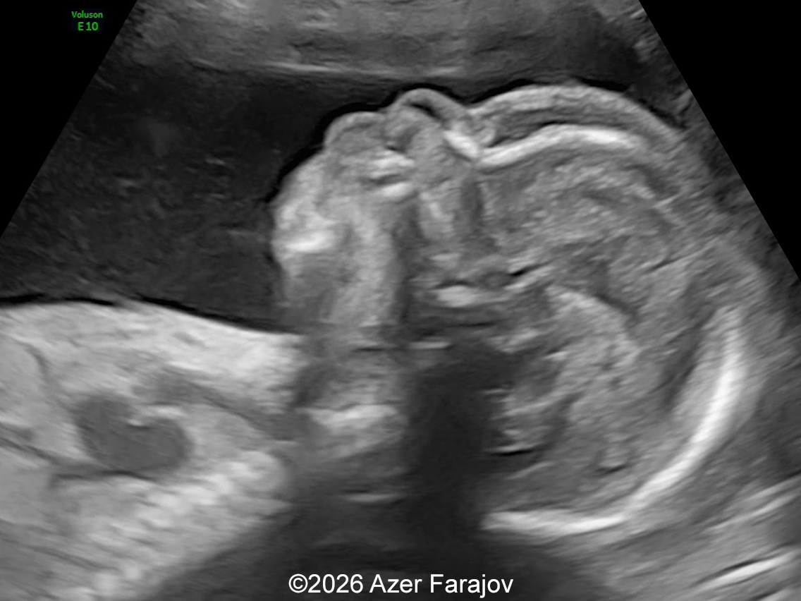
A 22-year-old primigravida presented for a routine ultrasound examination at 19 weeks and 6 days of gestation. The ultrasonographic evaluation revealed the following findings:
View the Answer Hide the Answer
Answer
We present a case of Congenital intrahepatic aorto-portal shunt (fistula).
Sonography demonstrated fetal skin edema, cardiomegaly with dilation of the ascending aorta and main pulmonary artery and marked dilatation of the porto-hepatic vascular system. Color Doppler imaging revealed a prominent vessel arising from the descending aorta and draining directly into the portal venous circulation, consistent with a congenital intrahepatic aorto-portal shunt. These findings suggested a high-flow arteriovenous communication with secondary fetal cardiac volume overload.
After multidisciplinary counseling regarding the prognosis, the parents opted for termination of pregnancy.
Discussion
Arteriovenous fistulas are abnormal communications that allow blood to shunt directly from an artery to a vein. Although they predominantly involve the peripheral vascular system, they may affect any organ or system in the body [1]. Histologically, arteriovenous fistulas consist of dysplastic arteries and veins that bypass the normal capillary bed and are not associated with soft tissue masses. This can lead to complications such as heart failure, anemia, embolism, and bleeding [2]. The diagnostic criteria on color and pulsed‑wave Doppler ultrasound include low‑ and high‑resistance flow patterns in the supplying artery, high‑velocity arterialized waveform in the draining vein, and turbulent, high‑velocity flow at the arteriovenous junction. These vascular lesions are uncommon, and in fetal life they have been described mainly in cranial structures such as the vein of Galen aneurysmal malformation, dural sinus malformation, and pial arteriovenous fistula. In the thoracic cavity, pulmonary arteriovenous malformations have occasionally been reported. Other fetal arteriovenous fistulas occur in the abdomen, and include intrahepatic arteriovenous fistulas, aorta–porto–umbilical vein fistulas, and distal abdominal aortocaval fistulas. These may arise in association with highly vascularized tumors such as sacrococcygeal teratomas, placental chorangiomas, and, less frequently, hepatoblastomas. In the prenatal period, the most frequent clinical manifestation is congestive heart failure secondary to the hemodynamic overload generated by the fistula.
Congenital arteriovenous shunts of the liver include hepatoportal arteriovenous shunts, hepatohepatic arteriovenous shunts, and portocaval anastomoses, with mixed forms combining more than one type [3,4]. These vascular anomalies occur in fewer than 1 in 100,000 live births and may lead to postnatal complications such as portal hypertension and high‑output cardiac failure [2]. Although most cases are sporadic, associations with chromosomal abnormalities such as trisomy 21 [5] and trisomy‑17 mosaicism [6], as well as with vascular malformation syndromes including hereditary hemorrhagic telangiectasia (Osler–Weber–Rendu disease) [7] and RASA1‑related capillary malformation–arteriovenous malformation syndrome [8] have been described. Consequently, detailed anatomic survey and consideration of genetic testing are reasonable.
Congenital aorto-portal or intrahepatic arterio-portal shunts represent a rare subtype of fetal abdominal arteriovenous malformations in which systemic arterial blood communicates directly with the portal or porto-umbilical venous system. The condition is described in the literature under various terms such as intrahepatic arterio-portal fistula and hepatic arterioportal shunt, in addition to other broader terms such as congenital abdominal/hepatic arteriovenous fistula. Despite differences in terminology, hepatohepatic arteriovenous shunts, portocaval anastomoses, and high‑flow hepatoportal arteriovenous shunts share a common pathophysiological mechanism: a low‑resistance, high‑flow vascular connection that increases venous return and results in high‑output cardiac physiology [3].
Hemodynamically, the lesion diverts a large volume of circulating blood (‘steal phenomenon') [2]. Blood is diverted from the aorta or hepatic arterial system into the portal circulation, reducing systemic vascular resistance while markedly increasing preload. The fetal heart compensates by augmenting cardiac output, which may progressively result in cardiomegaly, dilation of the great vessels, atrioventricular valve regurgitation, and ultimately congestive heart failure or hydrops fetalis [5,9]. Similar to other fetal high-flow vascular shunts, the cardiovascular manifestations are often the first diagnostic clue [10]. Dilatation of the pulmonary artery and ascending aorta most likely reflects chronically increased flow rather than primary structural cardiac disease.
Prenatal sonography typically reveals an intrahepatic vascular region without an associated solid mass; a key feature that helps distinguish this entity from hepatic tumors [11,12]. Accurate diagnosis requires identification of both the arterial feeder and the venous drainage pathway. Color Doppler demonstrates turbulent, high‑velocity flow with aliasing, whereas spectral Doppler may show arterialized pulsatile waveforms within the portal venous branches. Portal veins often appear dilated and pulsatile, and the draining hepatic or umbilical veins may exhibit increased flow velocities. These Doppler findings—arterialization of venous flow, demodulation with elevated diastolic velocities in the arterial waveform, and reversed flow within the drainage portal vein—are considered characteristic and enable reliable prenatal detection [13]. The use of 3D imaging, including multiplanar reconstruction and HD‑flow combined with STIC, further facilitates delineation of the relationship between the hepatic artery and the umbilico‑portal venous system, while also improving the assessment of potential fetal congenital heart defects [10,14].
Clinical presentation depends on the size and drainage pattern of the shunt. A larger fistula size (ratio of fistula diameter to umbilical vein diameter > 0.65 or ratio of fistula diameter to aorta > 0.9) increases the risk of heart failure [10]. Arterio-venous communications draining directly into hepatic or umbilical veins tend to produce earlier and more severe fetal cardiac compromise. In contrast, predominantly arterio-portal shunts may present postnatally with portal hypertension, hepatomegaly, ascites, or gastrointestinal bleeding [15]. Therefore, both cardiac and hepatic consequences should be anticipated.
Perinatal outcome largely depends on the degree of cardiac decompensation. Serial fetal echocardiography is recommended to monitor cardiac size, ventricular function, valve regurgitation, and signs of hydrops. Some small fistulas may go undetected during prenatal ultrasound, while others may close spontaneously following delivery [10]. Prenatal management strategies vary and include intrauterine corticosteroid therapy, maternal digoxin administration, and prophylactic use of steroids and propranolol [11, 12]. Delivery should be planned in a tertiary care center with pediatric cardiology and interventional radiology expertise. Postnatally, endovascular embolization is currently the preferred treatment and can be lifesaving, whereas surgical hepatic artery ligation, partial hepatic resection, and liver transplantation are reserved for selected cases [2,16,17].
The differential diagnosis includes congenital hepatic hemangioma or hemangioendothelioma with secondary shunting, congenital extrahepatic porto-systemic venous shunts (Abernethy malformation), and other causes of fetal high-output cardiac failure. The absence of a solid hepatic mass and the demonstration of a direct arterial feeder favor a primary arterio-portal shunt [18]. Other differential diagnosis is umbilico-portosystemic shunts, which consist in abnormal communication between one or several portal vein(s) and one or several hepatic vein(s). In these cases, the portal vein flows in a normal direction and is purely venous on pulsed Doppler [13].
In summary, congenital aorto-portal shunt is an uncommon but important cause of fetal cardiomegaly and high-output heart failure. Recognition of characteristic intrahepatic Doppler findings together with signs of cardiac volume overload enables early diagnosis, appropriate counseling, and coordinated perinatal management.
References
- González SB, Busquets JC, Figueiras RG, et al. Imaging arteriovenous fistulas. AJR Am J Roentgenol. 2009 Nov;193(5):1425-1433.
- Ungureanu A, Nagy RD, Constantin C, et al. Prenatal Diagnosis and Prognosis of Abdominal Arteriovenous Fistulae: A Comprehensive Case Series and Systematic Review. Diagnostics (Basel). 2024 Apr 17;14(8):826.
- Tseng JJ, Chou MM, Lee YH, Ho ES. Prenatal diagnosis of intrahepatic arteriovenous shunts. Ultrasound Obstet Gynecol. 2000 May;15(5):441-444.
- Zhou J, Zhou Q, Peng Q, Zeng S. Prenatal Diagnosis of Aorta-Porto-Umbilical Vein Fistulas with Left-Sided Inferior Vena Cava. AJP Rep. 2016 Mar;6(1):e59-61.
- Hartung J, Chaoui R, Kalache K, et al. Prenatal diagnosis of intrahepatic communications of the umbilical vein with atypical arteries (A-V fistulae) in two cases of trisomy 21 using color Doppler ultrasound. Ultrasound Obstet Gynecol. 2000 Sep;16(3):271-274.
- Iliescu DG, Ruican D, Nagy R, Burada F. Aorta-umbilical vein fistulae in fetus with trisomy-17 mosaicism. Ultrasound Obstet Gynecol. 2020 Mar;55(3):419-421.
- Saleh M, Miron I, Al-Rukban H, et al. Prenatal presentation of hereditary hemorrhagic telangiectasia - a report of two sibs. Prenat Diagn. 2016 Sep;36(9):891-893.
- Coccia E, Valeri L, Zuntini R, et al. Prenatal Clinical Findings in RASA1-Related Capillary Malformation-Arteriovenous Malformation Syndrome. Genes (Basel). 2023 Feb 22;14(3):549.
- Mathews B, Namdeo S, Krishnan V. Prenatal Diagnosis of Aorta-Portal Vein-Umbilical Vein Anastomosis. J Med Ultrasound. 2019 May 13;27(3):165.
- Li TG, Lv L, Zhang HX. Prenatal Diagnosis of Abdominal Aorta-Hepatic Artery-Umbilical Vein Fistula Using High-Definition Flow Render Mode and Spatiotemporal Image Correlation. J Ultrasound Med. 2026 Jun;45(6):1307-1316.
- Demirci O, Celayir A. Prenatal diagnosis and treatment of intrahepatic arteriovenous fistulas: case reports and the literature review. J Matern Fetal Neonatal Med. 2022 Mar;35(5):837-845.
- Mejides AA, Adra AM, O'Sullivan MJ, Nicholas MC. Prenatal diagnosis and therapy for a fetal hepatic vascular malformation. Obstet Gynecol. 1995 May;85(5 Pt 2):850-853.
- Douhnai D, Tassin M, Sibiude J, et al. Prenatal diagnosis of intra hepatic arterio venous fistula: case report and review of the literature. J Matern Fetal Neonatal Med. 2019 Aug;32(15):2575-2578.
- Van Houdt M, van der Merwe J, Gewillig M, De Catte L. Prenatal 3D-ultrasound diagnosis of isolated intrahepatic portal-systemic shunt with intact ductus venosus: A case report and literature review. Radiol Case Rep. 2021 Mar 16;16(5):1173-1178.
- Gedikbasi A, Oztarhan K, Sahin B, et al. Multidisciplinary approach to congenital multiple arterio-porto-caval malformation: case report. Am J Perinatol. 2008 May;25(5):265-270.
- Gladkikh M, Parra DA. Successful embolization of a congenital intra-hepatic arterioportal fistula in a neonate with the MVP Microvascular Plug system (MVP-3Q). Radiol Case Rep. 2022 Jan 18;17(3):991-996.
- Ramachandran P, Shanmugam NP, Vij M, Rela M. Surgical management of hepatic arterioportal fistula in a neonate. Pediatr Surg Int. 2014 May;30(5):557-559.
- Weng X, Jiang J, Wen G. Ultrasonographic diagnosis and follow-up of a special type of giant fetal hepatic hemangioma. Pediatr Radiol. 2025 May;55(5):1024-1028.
Discussion Board
Winners

Dianna Heidinger United States Sonographer

Andrii Averianov Ukraine Physician

Nutan Thakur India Physician

Ivan Ivanov Russian Federation Physician

Eti Zetounie Israel Sonographer
Murat Cagan Turkey Physician
Ionut Valcea Romania Physician

Hien Nguyen Van Viet Nam Physician
Almaz Kinzyabulatov Russian Federation Physician

Rupal Sasani India Physician

Nguyễn Lê Hoàng Viet Nam Physician

Tetiana Ishchenko Ukraine Physician

Hana Habanova Slovakia Physician

Mert Eyupoglu Turkey Physician

Gulten Rafibeyli Azerbaijan Physician

Ayten Sadigova Azerbaijan Physician
Aynur Garibova Azerbaijan Physician

Nataliia Antonenko Ukraine Physician