Case of the Week #654
Shivam Imaging, Ahmedabad, Gujarat, India
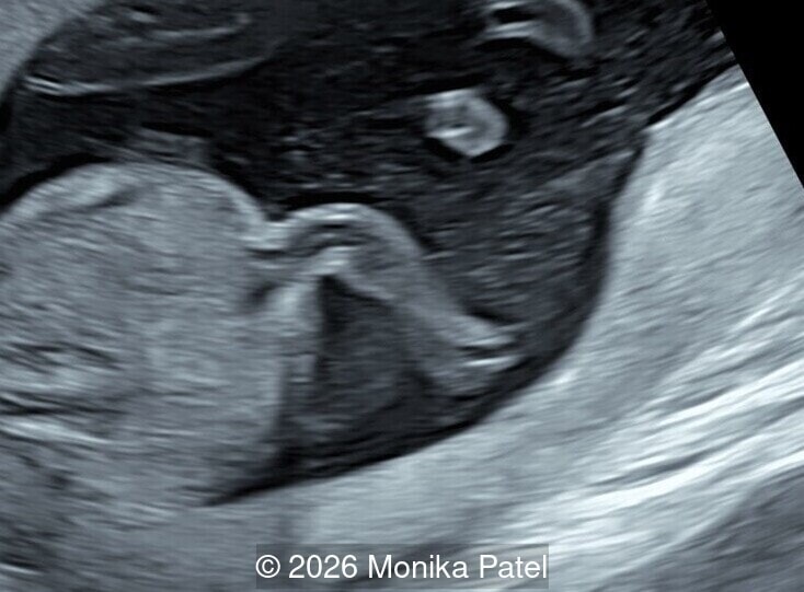
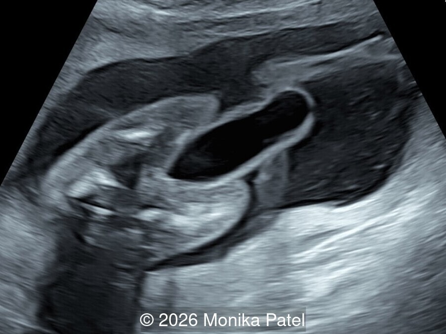
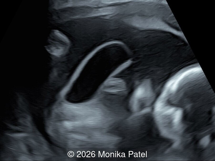
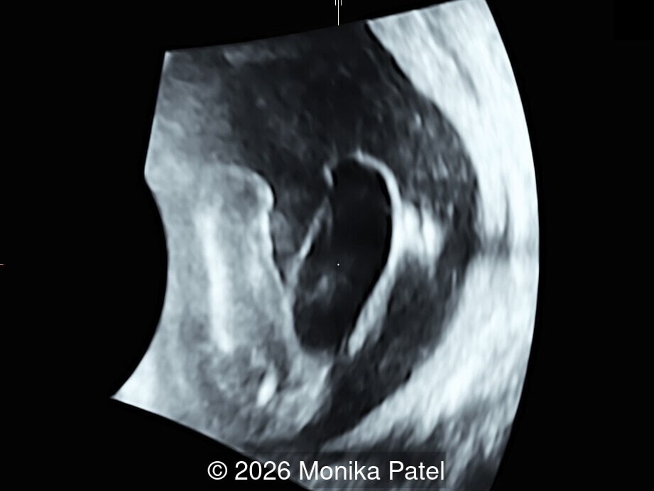
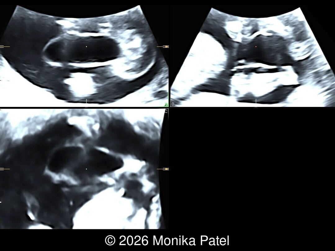
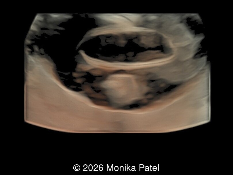
A G3P2 woman presented for routine antenatal scan. The following findings were seen.
View the Answer Hide the Answer
Answer
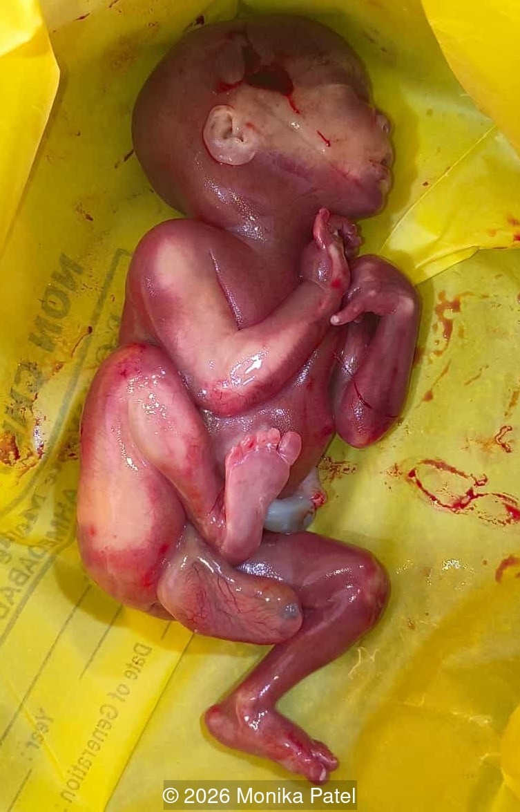
We present a case of congenital megalourethra. The patient elected to terminate the pregnancy. Exam of the abortus confirmed the diagnosis.
Our ultrasound images revealed a cystic tubular structure arising from pelvis without signs of lower urinary tract obstruction. Small intraabdominal bladder was seen. Our differential diagnosis included megalourethra, cloacal dysgenesis and rectal prolapse.
Discussion
First documented in 1949 [1] and termed “congenital megalourethra” in 1955 [2], the condition occurs due to faulty migration, differentiation, or development of the mesodermal tissues of the penis during the 7th week of gestation [3,4]. Because the urethra lacks the supportive structure of the surrounding erectile tissue, it balloons out into a cystic structure.
The condition is classified into two distinct clinical types [3]:
- Scaphoid Megalourethra (80% of cases): The corpus spongiosum is absent, but the corpora cavernosa remains intact. This creates a localized, boat-like ventral expansion of the urethra. It is the more common variant and tends to carry a more favorable prognosis, sometimes occurring as an isolated defect.
- Fusiform Megalourethra (20%): Both the corpus spongiosum and corpus cavernosum are deficient. This results in a circumferential, spindle-like dilation of the entire penis. This form is typically more severe and is frequently linked with other complex congenital anomalies, such as Prune Belly syndrome or VACTERL association [4-6].
The first prenatal diagnosis of megalourethra was in 1989 [7]. Megalourethra is typically diagnosed during the second trimester [4,6], though first-trimester diagnoses have been recorded [8]. On prenatal ultrasound, the defining characteristic is a prominent, fluid-filled cystic distention of the fetal penis, giving it an enlarged, balloon-like appearance [4,9]. Megalourethra is a functional urinary tract obstruction and frequently presents alongside other lower urinary tract obstruction markers, including [4,9]:
- Megacystis (52%): An enlarged, thick-walled urinary bladder.
- Keyhole Sign (12%): Produced by a dilated posterior urethra and an over-distended bladder.
- Hydronephrosis (58%) and Hydroureters (42%): Dilations of the kidneys and ureters caused by urinary stasis.
- Oligohydramnios (28%): Decreased amniotic fluid levels.
Diagnostic differentiation is critical; the dilated urethra can sometimes be mistakenly visualized as the umbilical cord [6]. Careful examination of the perineum and fetal genitalia is required.
The perinatal prognosis of fetal megalourethra is highly variable and depends on two main factors: the presence of associated syndromic malformations and the severity of renal damage. While isolated scaphoid megalourethra can have a good postnatal prognosis with surgical correction, fusiform megalourethra carries a poorer prognosis. Prolonged urinary stasis and functional obstruction in utero can lead to irreversible renal insufficiency, end-stage renal disease, or pulmonary hypoplasia [6]. In a study reviewing 50 cases of fetal megalourethra, mortality rate is 30% [4]. Ultrasound findings of bilateral hydroureters, bilateral hydronephrosis, severe oligohydramnios are associated with poor prognosis [4]. Prenatal spontaneous resolution of megalourethra occurs in 10% of fetuses often with normal amniotic fluid volume and without bilateral hydroureter and hydronephrosis [4]. Postnatally, functional obstruction can lead to recurrent urinary tract infections, renal insufficiency, sepsis and death. Postnatal renal impairment can be up to 40% [4].
Management and counseling require a multidisciplinary approach involving obstetricians, fetal medicine specialists, pediatric urologists, and neonatologists.
- Intrauterine Intervention: If oligohydramnios or significant renal impairment is detected early, in utero interventions such as vesicoamniotic shunting or serial bladder aspirations may be employed to relieve pressure and encourage normal pulmonary development [4].
- Postnatal Care: Postnatal imaging, such as a Micturating Cystourethrogram, can be performed to evaluate the exact anatomy and function of the urinary tract. The primary surgical treatment is a reduction urethroplasty to repair the urethra, restore a normal urinary stream, and improve the cosmetic appearance of the penis.
Congenital megalourethra is a rare urogenital anomaly requiring a high index of clinical suspicion during routine fetal anomaly scans. Prompt prenatal diagnosis enables vital counseling regarding the spectrum of the disease and facilitates early postnatal or intrauterine surgical intervention, which ultimately optimizes long-term renal and urological outcomes.
References
- Obrinsky W. Agenesis of abdominal muscles with associated malformation of the genitourinary tract; a clinical syndrome. Am J Dis Child (1911). 1949 Mar;77(3):362-73.
- Nesbitt TE. Congenital megalourethra. J Urol. 1955 May;73(5):839-42.
- Dorairajan T. Defects of spongy tissue and congenital diverticula of the penile urethra. Australian and New Zealand Journal of Surgery. 1962;32:209–214.
- Moaddab A, Sananes N, Hernandez-Ruano S, et al. Prenatal Diagnosis and Perinatal Outcomes of Congenital Megalourethra: A Multicenter Cohort Study and Systematic Review of the Literature. J Ultrasound Med. 2015 Nov;34(11):2057-64.
- Ardiet E, Houfflin-Debarge V, Besson R, et al. Prenatal diagnosis of congenital megalourethra associated with VACTERL sequence in twin pregnancy: favorable postnatal outcome. Ultrasound Obstet Gynecol 2003; 21:619–625.
- Sepulveda W, Elorza C, Gutierrez J, et al. Congenital megalourethra: outcome after prenatal diagnosis in a series of 4 cases. J Ultrasound Med. 2005 Sep;24(9):1303-8.
- Benacerraf BR, Saltzman DH, Mandell J. Sonographic diagnosis of abnormal fetal genitalia. J Ultrasound Med. 1989 Nov;8(11):613-7
- Lam YH, Tang MHY. Sonographic diagnosis of congenital megalourethra at 13 weeks’ gestation. Ultrasound Obstet Gynecol 2000; 16:585–586.
- Anh DD, Nguyen HT, Meagher S, et al. Prenatal diagnosis of congenital megalourethra in the second trimester of pregnancy. J Ultrason. 2019 Dec 31;19(79):302–304.
Discussion Board
Winners

Azar Farajov Azerbaijan Physician

Andrii Averianov Ukraine Physician

Nutan Thakur India Physician

Vladimir Lemaire United States Physician

Ivan Ivanov Russian Federation Physician

carlos lopez Venezuela Physician

CHARLES SARGOUNAME India Physician

Amparo Gimeno Spain Physician

Elena Andreeva Russian Federation Physician

Muradiye YILDIRIM Turkey Physician

Eti Zetounie Israel Sonographer

Deval Shah India Physician

Murat Cagan Turkey Physician
Siddhesh Rajiwade India Maternal fetal medicine specialist

Büşra Cambaztepe Turkey Physician

Ionut Valcea Romania Physician

Hien Nguyen Van Viet Nam Physician

Anette Beverdam Netherlands Sonographer

Annette Reuss Germany Physician

shruti Agarwal India Physician

Vu The Anh Viet Nam Physician
Rupal Sasani India Physician

Petra Majer Barboríková Slovakia Physician

Nguyễn Lê Hoàng Viet Nam Physician

Denys Saitarly Israel Physician

Tetiana Ishchenko Ukraine Physician
Costin Radu Lucian Romania Physician

Hana Habanova Slovakia Physician
Shamal Patel India Radiologist

DR RAJESH KAMBLE India Physician

Ashlea Hardin United States Sonographer

Ali Ozgur Ersoy Turkey Physician

ZHANNA Kurmangaliyeva Kazakhstan Physician

Mukesh Kannan India Consultant radiologist
Albert Guarque Rus Spain Physician
ANDRES ARENCIBIA MOLINA United States Physician

Maria Bulanova Russian Federation Physician

Mert Eyupoglu Turkey Physician

Gulten Rafibeyli Azerbaijan Physician

Ayten Sadigova Azerbaijan Physician

Aynur Garibova Azerbaijan Physician
Frederika Grochalova Slovakia Physician
Yaron Zalel Israel Physician
Sonu Kumar Batham India Physician
Nurten Asmalı çilek Turkey Physician
Nisa Hasanova Azerbaijan Physician