Case of the Week #653
(1) Israel, Meir Medical Center; (2) Centro Médico Recoletas, Valladolid, Spain
19-year-old primigravid woman with non-contributive medical history presented at 24 weeks for routine second trimester anomaly scan.
View the Answer Hide the Answer
Answer
We present a case of Crossed Fused Renal Ectopia. The diagnosis was confirmed on postnatal ultrasound.
Our ultrasound images demonstrated the following findings:
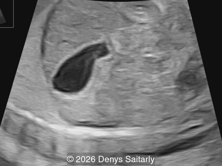
- Ultrasound image 1 and videos 1 and 2 demonstrate an empty left renal fossa and an inferiorly elongated left adrenal gland ("lying-down adrenal sign").
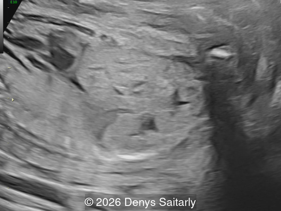
- Image 2 and video 3 show left-to-right crossed fused renal ectopia, with the ectopic kidney located inferior to the orthotopic right kidney (inferior crossed ectopia).
Discussion
Congenital renal anomalies in position and fusion arise from disruption of the normal embryologic migration of the kidneys. Between the sixth and ninth gestational weeks, the metanephros normally ascends from the pelvis to the retroperitoneal renal fossa in the lumbar region, undergoing a 90‑degree medial rotation along its longitudinal axis during this process [1]. Failure of the mature kidney to reach its usual location in the renal fossa results in renal ectopia, which may occur in several positions: pelvic, iliac, abdominal, thoracic, or contralateral/crossed [2,3].
Simple renal ectopia refers to a kidney located on the correct side of the body but in an abnormal position, either unilaterally or bilaterally. The most common form is the pelvic kidney, situated opposite the sacrum and below the aortic bifurcation. Kidneys that cross the midline are termed crossed renal ectopia, and describes a kidney located on the side opposite to its ureteral insertion into the bladder. Crossed renal ectopia may occur with or without fusion to the contralateral kidney.
Beyond positional abnormalities, shape and fusion anomalies include not only crossed renal ectopia with fusion but also horseshoe kidney. Horseshoe kidney consists of two vertically oriented renal masses connected at their lower poles by a parenchymal or fibrous isthmus that crosses the midline. Crossed fused renal ectopia is the second most common renal fusion anomaly after horseshoe kidney, with a reported incidence of 1 in 1,000 to 1 in 7,500 live births and a male predominance [4,5]. The majority of cases (79%) are detected antenatally, typically during routine second-trimester anatomic surveys [6]. Its etiology remains uncertain. The most widely accepted explanation is the “mechanical theory”, which proposes that an altered course of the umbilical arteries creates a path of least resistance that directs one metanephric unit toward the opposite side, facilitating fusion. Other proposed mechanisms include abnormal rotation of the caudal embryo, as well as the “ureteral theory,” in which the ureteral bud crosses the midline and induces differentiation of the contralateral metanephric blastema while the ipsilateral metanephros regresses [5,7].
McDonald and McClellan [8] classified crossed renal ectopia into four major categories describing the presence or absence of fusion and whether the anomaly is solitary or bilateral, then six additional subtypes based on the specific fusion pattern. Left‑to‑right ectopia accounts for approximately two‑thirds of cases, although proportions vary across published series [6,7]. Renal fusion occurs in about 90% of cases, and in at least 80% of these the crossed ectopic kidney lies inferior to the orthotopic kidney, with its upper pole fused to the lower pole of the normally positioned kidney (inferior ectopia). Additional morphological variants include sigmoid or S‑shaped, lump, and L‑shaped kidneys, as well as rare forms such as disk kidney and superior ectopia [6]. The ectopic kidney is often smaller than the orthotopic one, and its ureter crosses the pelvis to insert normally into the bladder [6]. The orthotopic kidney frequently exhibits anomalous vascularization, with multiple renal arteries arising at different levels from the aorta. The crossed ectopic kidney is typically supplied by one or more branches originating from the aorta or the common iliac artery on the side where it is located [9].
Failure to visualize a fetal kidney in its usual location—an “empty renal fossa”—is often interpreted as renal agenesis; however, careful examination may reveal an ectopic kidney [10-12]. Normal amniotic fluid volume and a visible fetal bladder provide reassurance that at least one functioning kidney is present. A pelvic kidney may be difficult to distinguish from adjacent structures, particularly in the second trimester when perinephric fat is limited [13]. When ectopia is suspected on a transverse image, sagittal and coronal views of the lower fetal abdomen and pelvis can assist in identifying ectopic renal tissue. Associated hydronephrosis or other renal anomalies may facilitate detection [14]. Color Doppler is essential for confirming the diagnosis by demonstrating the aberrant vascular supply to the ectopic kidney [5,15].
The adrenal gland arises from the mesodermal ridge in the thoracolumbar region, with contributions from neural crest cells [16]. Consequently, its position is not affected by renal migration abnormalities. When a kidney is absent from the renal fossa, the adrenal gland, which is normally located superior to the kidney and characterized by a hypoechoic cortex and hyperechoic medulla, appears flattened and vertically oriented and occupies the empty fossa. This is known as the “lying‑down adrenal sign” [17,18].
The first prenatal diagnosis of crossed renal ectopia was reported by Greenblatt et al. [19] in 1985. Since then, it has been described only sporadically, within broader series of renal ectopia [10-12], in combined pre‑ and postnatal studies [6,7], or as isolated cases or small dedicated series [14,15,20-22]. In some fetuses with an empty renal fossa, meticulous sonographic evaluation reveals the typical appearance of crossed renal ectopia: one kidney, usually the right, occupies its normal position, whereas the contralateral kidney lies inferior to it and is malrotated [3]. A cross‑fused ectopic kidney may mimic an enlarged solitary kidney when the contralateral kidney is not visualized; therefore, assessment of renal morphology and contour, together with identification of two echogenic collecting systems, is essential for correct diagnosis [10]. Because renal ascent is completed by the eighth gestational week, a fetal pelvic kidney can be detected as early as the first trimester using high‑frequency transvaginal probes [22].
Urinary tract anomalies occur in 24-41% of cases and include hydronephrosis (38%), vesicoureteral reflux (41%), double collecting systems, and ureteropelvic junction obstruction [6,15,23]. Extraurinary anomalies are present in approximately 40-48% of cases and can include cardiovascular, skeletal, genital and gastrointestinal abnormalities [6,15, 24]. Cardiac anomalies include ventricular septal defect and persistent left superior vena cava [6,15]. Genital anomalies include hypospadias and cryptorchidism [6]. Gastrointestinal abnormalities include anorectal malformation, which may occur in isolation or as part of the VACTERL association (Vertebral anomalies, Anal atresia, Cardiac malformations, Tracheoesophageal fistula, Renal anomalies, Limb abnormalities) [7,24]. Single umbilical artery has also been associated with crossed fused renal ectopia [15].
The sonographic identification of any form of renal ectopia should not modify obstetric management. Once the diagnosis is established, a meticulous ultrasound examination is required to exclude associated anomalies, followed by periodic sonographic surveillance throughout pregnancy [15].
Cross‑fused ectopia should not be misinterpreted as unilateral renal agenesis with compensatory hypertrophy of the contralateral kidney. Beyond this scenario, the main differential diagnosis is a renal tumor [19]. Several sonographic features help distinguish renal ectopia from a neoplastic mass: a normally positioned kidney will be present in the contralateral renal fossa; the characteristic echogenic central collecting system is absent in tumors; and the echotexture of a mass typically differs from that of normal renal parenchyma.
Postnatal evaluation includes renal-bladder ultrasound, which is usually sufficient for confirmation of the diagnosis. Additional imaging with voiding cystourethrography (VCUG) and dimercaptosuccinic acid (DMSA) scan may be indicated to detect vesicoureteral reflux and assess differential renal function [6]. Postnatal nephrological follow-up is recommended for all cases [15]
Most individuals with an ectopic kidney remain asymptomatic throughout life, with the condition often identified incidentally. However, abnormal renal position and/or aberrant ureteral course may predispose to complications such as hydronephrosis, vesicoureteral reflux, ureteropelvic junction obstruction, urinary stone formation, and infection [25-30]. Impaired renal function in the ectopic kidney occurs in approximately 34% of cases on postnatal DMSA scanning, though chronic renal failure is uncommon in isolated cases [6,31]. Associated urinary anomalies require postnatal surgical intervention in approximately one-third of affected patients [6]. The presence of extraurinary malformations with crossed fused renal ectopia carries higher morbidity risk. In one series, 14% of patients with extraurinary anomalies developed chronic kidney disease, with mortality occurring in those with severe associated malformations [6].
References
- Carlson BM. Urogenital system. In: Human Embryology and Developmental Biology. 6th ed. St. Louis (MO): Elsevier; 2019. p. 358‑390.e2.
- Shapiro E, Bauer SB, Chow JS. Anomalies of the upper urinary tract. In: Wein AJ, editor. Campbell‑Walsh Urology. 10th ed. Philadelphia (PA): Elsevier; 2012. p. 3123–3160.
- Bleich AT, Dashe JS. Abnormal kidney location. In: Copel JA, editor. Obstetric Imaging: Fetal Diagnosis and Care. 3rd ed. Philadelphia (PA): Elsevier; 2026. p. 387–391.e1.
- Glodny B, Petersen J, Hofmann KJ, et al. Kidney fusion anomalies revisited: clinical and radiological analysis of 209 cases of crossed fused ectopia and horseshoe kidney. BJU Int. 2009 Jan;103(2):224-235.
- Kubihal V, Razik A, Sharma S, Das CJ. Unveiling the confusion in renal fusion anomalies: role of imaging. Abdom Radiol (NY). 2021 Sep;46(9):4254-4265.
- Sarhan O, El Helaly A, Al Otay A, et al. Crossed fused renal ectopia: Diagnosis and prognosis as a single-center experience. J Pediatr Surg. 2021 Sep;56(9):1632-1637.
- Loganathan AK, Bal HS. Crossed fused renal ectopia in children: a review of clinical profile, surgical challenges, and outcome. J Pediatr Urol. 2019 Aug;15(4):315-321.
- McDonald JH, McClellan DS. Crossed renal ectopia. Am J Surg. 1957 Jun;93(6):995-1002.
- Rubinstein ZJ, Hertz M, Shahin N, Deutsch V. Crossed renal ectopia: angiographic findings in six cases. AJR Am J Roentgenol. 1976 May;126(5):1035-1038.
- Jeanty P, Romero R, Kepple D, et al. Prenatal diagnoses in unilateral empty renal fossa. J Ultrasound Med. 1990 Nov;9(11):651-654.
- Yuksel A, Batukan C. Sonographic findings of fetuses with an empty renal fossa and normal amniotic fluid volume. Fetal Diagn Ther. 2004 Nov-Dec;19(6):525-532.
- Chow JS, Benson CB, Lebowitz RL. The clinical significance of an empty renal fossa on prenatal sonography. J Ultrasound Med. 2005 Aug;24(8):1049-1054.
- Bowie JD, Rosenberg ER, Andreotti RF, Fields SI. The changing sonographic appearance of fetal kidneys during pregnancy. J Ultrasound Med. 1983 Nov;2(11):505-507.
- Chang PL, Mrazek-Pugh B, Blumenfeld YJ. Prenatal diagnosis of cross-fused renal ectopia: does color Doppler and 3-dimensional sonography help? J Ultrasound Med. 2011 Apr;30(4):578-580.
- Zajicek M, Perlman S, Dekel B, et al. Crossed ectopic kidney: prenatal diagnosis and postnatal follow-up. Prenat Diagn. 2017 Jul;37(7):712-715.
- Ross IL, Louw GJ. Embryological and molecular development of the adrenal glands. Clin Anat. 2015 Mar;28(2):235-242.
- Hoffman CK, Filly RA, Callen PW. The "lying down" adrenal sign: a sonographic indicator of renal agenesis or ectopia in fetuses and neonates. J Ultrasound Med. 1992 Oct;11(10):533-536.
- Majmudar A, Cohen HL. "Lying-Down" Adrenal Sign: There Are Exceptions to the Rule Among Fetuses and Neonates. J Ultrasound Med. 2017 Dec;36(12):2599-2603.
- Greenblatt AM, Beretsky I, Lankin DH, Phelan L. In utero diagnosis of crossed renal ectopia using high-resolution real-time ultrasound. J Ultrasound Med. 1985 Feb;4(2):105-107.
- Siegel RL, Rosenfeld DL, Leiman S. Complete regression of a multicystic dysplastic kidney in the setting of renal crossed fused ectopia. J Clin Ultrasound. 1992 Sep;20(7):466-469.
- Singhania S, Singhania A, Khan S, et al. Prenatal Diagnosis of Cross-fused Renal Ectopia: Still a Dilemma. Donald School J Ultrasound Obstet Gynecol 2017;11(3):225-226.
- Poenaru MO, Augustin FE, Vâlcea ID, et al. Ectopic Kidney, a Challenging First Trimester Diagnosis-Case Report and Literature Review. Life (Basel). 2024 Nov 12;14(11):1466.
- Inan C, Sayin C, Varol F. Prenatal diagnosis of ectopic kidney: Evaluation of characteristics, additional anomalies and urinary complications. Eur J Obstet Gynecol Reprod Biol. 2024 Sep:300:150-154.
- Rivard DJ, Milner WA, Garlick WB. Solitary crossed renal ectopia and its associated congenital anomalies. J Urol. 1978 Aug;120(2):241-242.
- Zheng Y, Wang Y, Ren S, Wang D. Crossed fused renal ectopia with hydronephrosis: A case report. Asian J Surg. 2023 Aug;46(8):3087-3088.
- Kesan K, Gupta R, Gupta A, et al. Solitary crossed renal ectopia with vesicoureteric reflux presenting with impaired renal function in a neonate. J Clin Neonatol. 2013 Jul;2(3):140-142.
- Gebreamlak AL, Alema KT, Kumsa ID, Molla MM. A case report on pyelovesicostomy of PUJO of pelvic crossed fused renal ectopia with single ureter. Int J Surg Case Rep. 2024 Feb;115:109212.
- Kramer SA, Kelalis PP. Ureteropelvic junction obstruction in children with renal ectopy. J Urol (Paris). 1984;90(5):331-336.
- Benabdallah W, Othmane MB, Bouassida K, et al. Crossed-fused renal ectopia with renal calculi and its management, a case report. Int J Surg Case Rep. 2023 May;106:108218.
- Jaiswal P, Talwar D, Acharya S, et al. Crossed Fused Renal Ectopia With Acute Pyelonephritis: A Case Report. Cureus. 2021 Nov 25;13(11):e19907.
- Agar BE, Gurgoze MK, Kara A. Clinical Significance and Prognosis of Renal Fusion Anomalies Detected by Tc-99m Dimercaptosuccinic Acid Scintigraphy. Clin Pediatr (Phila). 2026 Jun;65(5):682-688.
Discussion Board
Winners

Andrii Averianov Ukraine Physician

Alexandr Krasnov Ukraine Physician

Mayank Chowdhury India Physician

Vladimir Lemaire United States Physician

Boujemaa Oueslati Tunisia Physician
yaser hamidian Iran, Islamic Republic of Physician

Amparo Gimeno Spain Physician

Muradiye YILDIRIM Turkey Physician

ALBANA CEREKJA Italy Physician

Murat Cagan Turkey Physician
Nguyen Thac Viet Viet Nam Physician

Büşra Cambaztepe Turkey Physician

gholamreza azizi Iran, Islamic Republic of Physician

Ionut Valcea Romania Physician

Kathrine Montagne United States Sonographer

Anette Beverdam Netherlands Sonographer

Annette Reuss Germany Physician

CHERYL TURNER United States Sonographer

shay kevorkian Israel Physician

Sruthi Pydi India Physician

Eylem Eşsizoğlu Turkey Physician

Nguyễn Lê Hoàng Viet Nam Physician
Rajnikant Vasava India Physician

Denys Saitarly Israel Physician

Tetiana Ishchenko Ukraine Physician

Hana Habanova Slovakia Physician
Kelsey O'Brien United States Sonographer

Ashlea Hardin United States Sonographer

Mukesh Kannan India Consultant radiologist
Ngoc Bich Viet Nam Physician

MISCHEL BARTIE United States Sonographer

Mert Eyupoglu Turkey Physician

Gulten Rafibeyli Azerbaijan Physician

Ayten Sadigova Azerbaijan Physician
Aynur Garibova Azerbaijan Physician
Elnara Baghirova Azerbaijan Physician