Case of the Week #652
UT Southwestern Medical Center, Plano Texas, United States
25-year-old primigravida was referred to our facility at 26 weeks 4 days gestation for a detailed fetal anatomic survey. There were no pertinent maternal and obstetric history. Noninvasive prenatal testing showed a male fetus at low risk for chromosomal abnormalities.
View the Answer Hide the Answer
Answer
We present a case of isolated mild aortic stenosis, valvular type.
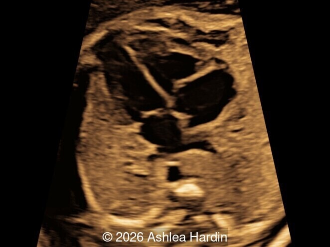
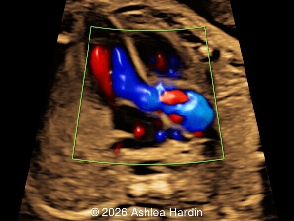
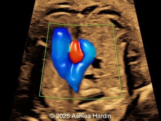
Our images revealed thickened aortic valve leaflets with dilation of the aortic root distal to the aortic valve. Turbulent antegrade flow can be seen with color doppler in the 5-chamber view, three vessel trachea view and the right ventricular outflow view.
The patient denied any further genetic testing, but accepted referral to pediatric cardiology. Pediatric cardiology performed an initial fetal echocardiogram at 27 weeks gestation that confirmed mild aortic stenosis with a peak velocity of 210cm/s, and a dilated ascending aorta measuring 0.75 cm with a Boston z-score of 3.52. A follow up echocardiogram at 31 weeks gestation showed stable findings.
Discussion
Aortic stenosis is defined as narrowing at the level of the aortic valve leading to an obstruction of left ventricular outflow tract. Aortic stenosis occurs in 3-6% of structural heart defects and is more common in males. There are several types of aortic stenosis, each named after the anatomical relation to the aortic valve: valvular, subvavular and supravalvular. Valvular aortic stenosis is the most common type diagnosed prenatally. Subvalvular and supravalvular are less commonly seen in utero, especially when isolated.
Mild valvular aortic valve stenosis may go undiagnosed in gestation due to a normal four-chamber view. Using the apical five-chamber view, a dilated aortic root distal to the aortic valve can be seen with thickened aortic valve leaflets. In this case, an abnormal five-chamber view shows dilation of the aortic root, which then can be seen extending into the three-vessel-trachea view. Using color Doppler, turbulent antegrade flow in the three-vessel-trachea view is demonstrated, which can also been seen in the high short axis view as the pulmonary artery wraps around the aorta.
Development of hypoplastic left heart syndrome due to left ventricular dysfunction is an indication of a worse prognosis in patients with aortic stenosis. The prognosis is generally good in those with mild aortic stenosis without signs hypoplastic left heart syndrome, .
Mild aortic stenosis is commonly associated with a bicuspid aortic valve. Bicuspid aortic valve alone is not commonly diagnosed in prenatal ultrasound. Differential diagnosis also includes transposition of the great arteries with a dilated root of the pulmonary artery, which could mimic aortic stenosis. Tetralogy of Fallot could also imitate aortic stenosis due to turbulent flow across the aortic valve.
References
Abuhamad, Alfred, and Rabih Chaoui. “Chapter 21: Aortic Stenosis and Bicuspid Aortic Valve.” A Practical Guide to Fetal Echocardiography Normal and Abnormal Hearts, Third ed., Wolters Kluwer, Philadelphia, PA, 2016, pp. 317–328.
Discussion Board
Winners

Azar Farajov Azerbaijan Physician

Dianna Heidinger United States Sonographer

paola quaresima Italy Physician

Andrii Averianov Ukraine Physician

Mayank Chowdhury India Physician

Nutan Thakur India Physician

Boujemaa Oueslati Tunisia Physician

CHARLES SARGOUNAME India Physician

Kimberly Delaney United States Sonographer
Olivia Ionescu United Kingdom Physician
Javier Ayala Spain Physician
yaser hamidian Iran, Islamic Republic of Physician

Amparo Gimeno Spain Physician

Elena Andreeva Russian Federation Physician

Eti Zetounie Israel Sonographer
Murat Cagan Turkey Physician

Eslam Adel ammar Egypt Physician
rahous karim United States

ANA PAULA PASSOS Brazil Physician
gholamreza azizi Iran, Islamic Republic of Physician
Ionut Valcea Romania Physician

Hien Nguyen Van Viet Nam Physician

Kathrine Montagne United States Sonographer
Almaz Kinzyabulatov Russian Federation Physician
Zuzana Briešková Slovakia Physician
Ann-Christin Dr. Sönnichsen Germany Physician

Anette Beverdam Netherlands Sonographer

shruti Agarwal India Physician

YULIA VISHNEVSKAYA Russian Federation Physician
Brindusa Ciobanu Romania Physician

shay kevorkian Israel Physician
Shelby Duncan United States Sonographer

Sruthi Pydi India Physician
Oanh Oanh Viet Nam Physician
Rohit Sanghani India Physician
Rupal Sasani India Physician
Joanna Głowska-Ciemny Poland Physician

Nguyễn Lê Hoàng Viet Nam Physician
Rajnikant Vasava India Physician

Denys Saitarly Israel Physician
Costin Radu Lucian Romania Physician

Hana Habanova Slovakia Physician
Kelsey O'Brien United States Sonographer

Văn Kiệt Võ Viet Nam Physician
sugunadevi Viswanathan India Physician

Ali Ozgur Ersoy Turkey Physician
Lucia Bobik Slovakia Physician

Viktorya Mkrtchyan Armenia Physician
ZHANNA Kurmangaliyeva Kazakhstan Physician
ERTAN KARAMAN Turkey Physician

Mukesh Kannan India Consultant radiologist
RAVINDRANATH MALLELA India RADIOLOGIST

Luu Anh Viet Nam Physician
Balpreet Duggal Trinidad and Tobago Physician
Vanessa Roach Scott Australia Sonographer
Bruna Eduarda Peres Maciel Castro Brazil Physician
Harpinder Singh India Physician
Anne Janke Germany Physician

Mert Eyupoglu Turkey Physician

Gulten Rafibeyli Azerbaijan Physician

Gulsum Mammadova Azerbaijan Physician
Aynur Garibova Azerbaijan Physician
Petra Mariacher Austria Physician
Elnara Baghirova Azerbaijan Physician

Nataliia Antonenko Ukraine Physician
Melissa Lin Australia Physician
Kushagra Mittal India Physician
Nashwa Hegazy Saudi Arabia Physician