Case of the Week #651
(1) Femicare, Center of prenatal ultrasonographic diagnostics, Martin, Slovakia; (2) Centro Médico Recoletas, Valladolid, Spain
27-year-old G2P1 with a non-contributory history presented to our office at 19 weeks gestation.
View the Answer Hide the Answer
Answer
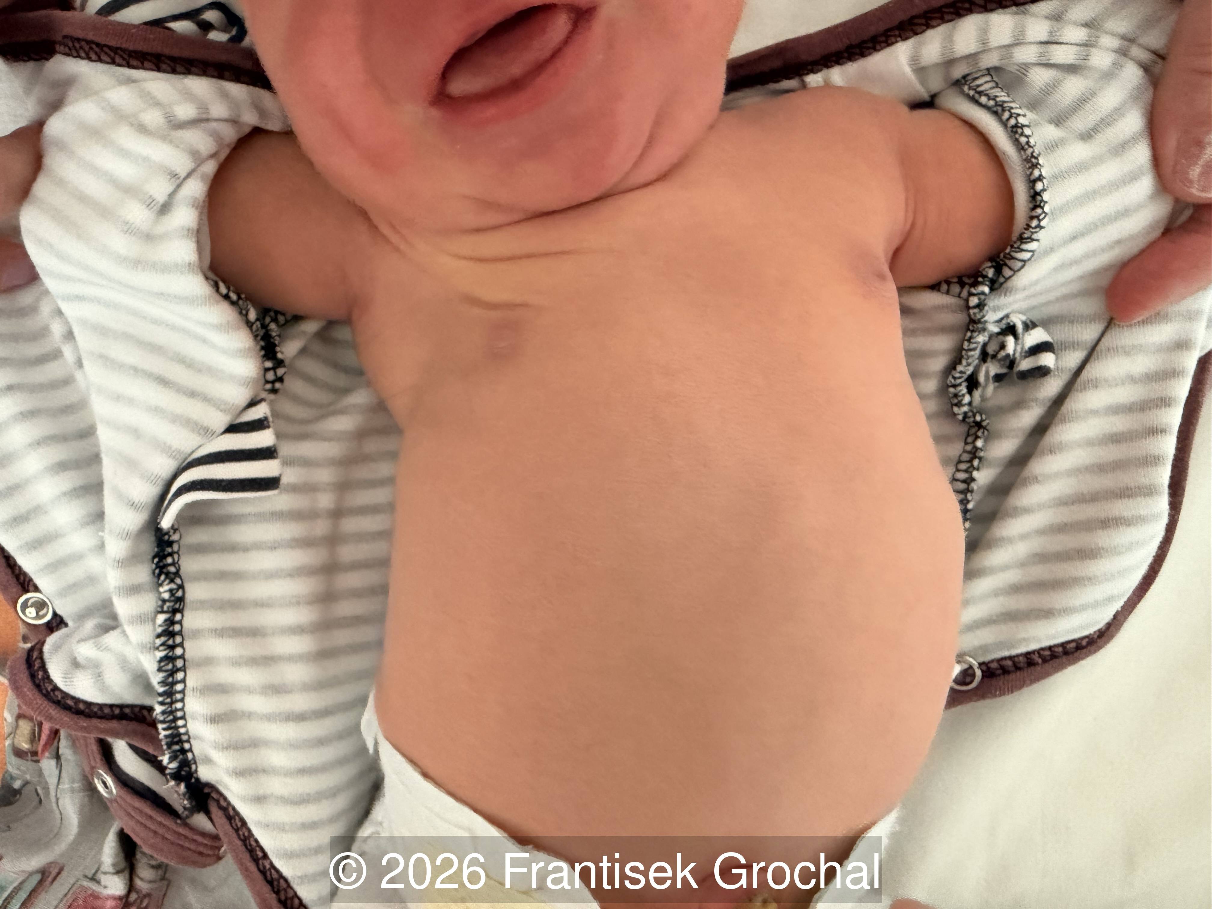
We present a case of Poland syndrome. The pregnancy resulted in a spontaneous term delivery at 39 weeks of a male neonate weighing 3700g and measuring 51 cm with Apgar scores of 9, 9, 9. On retrospective review of the videos, and with knowledge of the diagnosis, the absence of the right pectoralis major muscle appears appreciable.
Discussion
A systematic assessment of the fetal thoracic wall, although often underappreciated, is an essential component of prenatal ultrasound, as it allows early identification of structural abnormalities such as agenesis or hypoplasia of the pectoralis muscles, rib defects, sternal clefts, and asymmetric thoracic development. Recognition of these findings is particularly relevant for the diagnosis of conditions in which thoracic morphology is a defining feature, including Poland syndrome, Jeune syndrome (asphyxiating thoracic dystrophy), Jarcho–Levin syndrome, skeletal dysplasias, and other disorders encompassed within the broader pediatric concept of “thoracic insufficiency syndrome”. Thoracic size and contour provide indirect but informative markers of pulmonary development, especially regarding the risk of pulmonary hypoplasia in lethal skeletal dysplasias. Furthermore, correlating thoracic abnormalities with anomalies of the upper limb or shoulder girdle enhances diagnostic accuracy in syndromic presentations where these features coexist. Although limited, evaluation of fetal thoracic excursion may also offer supportive clues to neuromuscular dysfunction or mechanical restriction.
Poland syndrome (also referred to as Poland anomaly or Poland sequence) is a rare congenital musculoskeletal condition characterized by hypoplasia or aplasia of the pectoral muscles, mammary hypoplasia, and variably associated ipsilateral upper‑limb malformations [1]. The condition was formally described in 1841 by Sir Alfred Poland [2] in Guy’s Hospital Reports, following the autopsy of a 27‑year‑old convict who exhibited absence of the pectoral muscles together with ipsilateral syndactyly. However, an earlier autopsy report published in 1839 by Froriep [3], titled “Observation of a Case of Absence of the Breast”, documented a woman with amastia, absence of the third and fourth ribs, and agenesis of the pectoralis major, pectoralis minor, intercostal muscles, and portions of the serratus muscle. Because of this prior description, and given that the association between absence of the pectoralis major and ipsilateral syndactyly was not recognized as a unified entity until 1895–1900, the term Poland syndrome—proposed in 1962 by Clarkson [4], a hand surgeon also at Guy’s Hospital—illustrates the often-misleading nature of eponymous disease nomenclature [5].
Because of its variable clinical presentation, Poland syndrome is often not recognized at birth. Owing to underreporting, the true incidence of the condition remains uncertain; however, current estimates suggest a frequency of approximately 1:30,000 to 1:90,000 live births [6,7]. Milder forms may go unnoticed until late adolescence or puberty, when chest asymmetry becomes more apparent. The right side is affected about twice as often as the left, and although the anomaly is two to three times more common in boys than in girls, asymmetric breast development tends to be more readily detected in female patients [6]. Most cases occur sporadically, but familial occurrences with intrafamilial variability have been reported [8-10], leading some authors to propose an autosomal dominant inheritance with low penetrance or a multifactorial model [8]. The marked clinical variability, together with the bilateral manifestations described in some individuals, is more consistently explained by a paradominant mechanism, in which a germline mutation remains clinically silent unless a postzygotic “second hit” induces mosaic loss of the normal allele, generating a cell clone that is either homozygous or hemizygous for the mutation [11,12]. In the absence of a family history of Poland sequence, the recurrence risk is considered to be considerably less than 1% [8].
Several etiological hypotheses have been proposed, although the vascular disruption theory remains the most widely accepted pathogenic mechanism. According to this model, an obstruction of the subclavian artery proximal to the origin of the internal thoracic artery but distal to the vertebral artery around the sixth week of gestation leads to reduced blood flow to the distal upper limb and pectoral region, resulting in the localized tissue loss characteristic of Poland syndrome. The concept of the subclavian artery supply disruption sequence (SASDS), introduced by Bavinck and Weaver [13], encompasses these embryonic vascular disturbances and proposes a shared pathogenic pathway for Poland, Klippel-Feil, and Möbius syndromes, as well as for isolated terminal transverse limb defects and Sprengel deformity. Möbius syndrome is defined by unilateral or bilateral sixth‑ and seventh‑cranial‑nerve palsies, leading to abducens paresis and facial weakness. Approximately 15-20% of cases show an association with Poland syndrome, giving rise to the overlapping Möbius-Poland phenotype [14].
Although the original definition of Poland syndrome required the coexistence of thoracic and upper‑limb anomalies, it is now widely accepted that the only mandatory diagnostic criterion is total or partial agenesis of the pectoralis major muscle. The spectrum of thoracic defects is broad, ranging from subtle breast hypoplasia to amastia, as well as hypoplasia and reduced pigmentation of the nipple–areola complex, rib and cartilage hypoplasia, chest‑wall depression, sternal anomalies, absence of axillary hair, and diminished subcutaneous fat [1]. Most cases of Poland sequence involve the right side, and only a minority present with rib hypoplasia. However, when the defect is left‑sided, rib anomalies are more frequent and are often accompanied by isolated dextrocardia, which typically occurs only when there is partial agenesis of two or more ribs [15]. The classic hand deformity includes syndactyly and a variable degree of brachydactyly, with marked hypoplasia or aplasia of the middle phalanges (brachysyndactyly). Nevertheless, upper‑limb malformations may be absent or, at the opposite extreme, as severe as a phocomelia‑like deficiency [16]. Additional associated anomalies have been reported in some cases, including vertebral defects, renal aplasia or hypoplasia, and lower‑limb abnormalities. Based on their experience with 245 patients with Poland syndrome, Romanini et al. [17] proposed a phenotypic classification grounded in the most frequent abnormalities: Type 1, or minimal form, when the pectoral muscle defect is isolated; Type 2, or partial forms, when it is associated with either a rib anomaly or an upper‑limb defect; and Type 3, or complete form, when the pectoral muscle defect is associated with both.
When prenatal ultrasound reveals unilateral limb abnormalities together with chest asymmetry, a diagnosis of Poland sequence should be considered. Four cases of prenatal diagnosis have been reported in the literature [18-21], although a truly prenatal diagnosis was achieved in only two—those described by Paladini and Nguyen. In both reports, the initial finding was chest‑wall asymmetry, which in the latter case was explicitly attributed to absence of the pectoralis major muscle. Additional anomalies present in both fetuses facilitated diagnostic recognition: upper‑limb defects in the first, which are common in Poland syndrome, and brain anomalies in the second, characteristic of Poland–Möbius syndrome. The remaining two cases correspond to postnatal diagnoses. In the case reported by Sepúlveda, an anterior chest‑wall deformity was present in a fetus with cardiac dextroposition, a finding typically associated with agenesis of the left pectoralis major muscle, but it was not identified on third‑trimester ultrasound or MRI. In the case described by Berdel, right‑hand aplasia was noted prenatally, but Poland syndrome was diagnosed only at three months of age, when absence of the right pectoralis major muscle was confirmed. In addition to potential findings on B‑mode ultrasound, three‑dimensional ultrasound can assist in characterizing the limb defect, and Doppler assessment of ipsilateral subclavian artery flow may also provide supportive evidence for the diagnosis.
Pectoral muscle hypoplasia is not specific to Poland syndrome and may also occur in syndromes with a defined genetic basis that feature hand, pectoral, and other associated anomalies, such as Holt–Oram syndrome, Duane radial ray syndrome, CHILD syndrome, craniofacial microsomia, and ulnar‑mammary (Levy–Hollister) syndrome. Therefore, in patients with an atypical phenotype, such as congenital heart disease, ocular or otological manifestations, alternative diagnoses or syndromic conditions should be considered before establishing a diagnosis of Poland syndrome. Misclassification may have important implications for the patient and their family regarding inheritance patterns and the potential presence of additional anomalies [22].
References
- Hashim EAA, Quek BH, Chandran S. A narrative review of Poland's syndrome: theories of its genesis, evolution and its diagnosis and treatment. Transl Pediatr. 2021 Apr;10(4):1008-1019.
- Poland A. Deficiency of the pectoral muscles. Guys Hosp Rep. 1841;6:191-193.
- Froriep R. Beobachtung eines Falles von Mangel der Brustdrüse: Notizen aus dem Gebiete der Natur- und Heilkunde. 1839;10:9–14.
- Clarkson P. Poland's syndactyly. Guys Hosp Rep. 1962;111:335-346.
- Ram AN, Chung KC. Poland's syndrome: current thoughts in the setting of a controversy. Plast Reconstr Surg. 2009 Mar;123(3):949-953.
- McGillivray BC, Lowry RB. Poland syndrome in British Columbia: incidence and reproductive experience of affected persons. Am J Med Genet. 1977;1(1):65-74.
- Czeizel A, Vitéz M, Lenz W. Birth prevalence of Poland sequence and proportion of its familial cases. Am J Med Genet. 1990 Aug;36(4):524.
- David TJ. Familial Poland anomaly. J Med Genet. 1982 Aug;19(4):293-296.
- Darian VB, Argenta LC, Pasyk KA. Familial Poland's syndrome. Ann Plast Surg. 1989 Dec;23(6):531-537.
- Shalev SA, Hall JG. Poland anomaly--report of an unusual family. Am J Med Genet A. 2003 Apr 15;118A(2):180-183.
- Happle R. Poland anomaly may be explained as a paradominant trait. Am J Med Genet. 1999 Dec 3;87(4):364-365.
- Baban A, Torre M, Bianca S, et al. Poland syndrome with bilateral features: case description with review of the literature. Am J Med Genet A. 2009 Jul;149A(7):1597-1602.
- Bavinck JN, Weaver DD. Subclavian artery supply disruption sequence: hypothesis of a vascular etiology for Poland, Klippel-Feil, and Möbius anomalies. Am J Med Genet. 1986 Apr;23(4):903-918.
- Agarwal R, Kumar M, Vasudeva A, et al. Möbius Syndrome With Possible Poland Syndrome Overlap: A Case Report. Cureus. 2025 Mar 2;17(3):e79916.
- Torre M, Baban A, Buluggiu A, et al. Dextrocardia in patients with Poland syndrome: phenotypic characterization provides insight into the pathogenesis. J Thorac Cardiovasc Surg. 2010 May;139(5):1177-1182.
- Al-Qattan MM. Classification of hand anomalies in Poland's syndrome. Br J Plast Surg. 2001 Mar;54(2):132-136.
- Romanini MV, Calevo MG, Puliti A, et al. Poland syndrome: A proposed classification system and perspectives on diagnosis and treatment. Semin Pediatr Surg. 2018 Jun;27(3):189-199.
- Paladini D, D'Armiento MR, Martinelli P. Prenatal ultrasound diagnosis of Poland syndrome. Obstet Gynecol. 2004 Nov;104(5 Pt 2):1156-1159.
- Sepulveda W. Poland syndrome: a rare cause of cardiac dextroposition in the fetus. Prenat Diagn. 2009 Sep;29(9):903-905.
- Berdel AL, Henrich W. Antenatal sonographic features of Poland syndrome on 2- and 3-dimensional sonography. J Ultrasound Med. 2010 Apr;29(4):679-680.
- Nguyen GV, Goncalves LF, Vaughn J, et al. Prenatal diagnosis of Poland-Möbius syndrome by multimodality fetal imaging. Pediatr Radiol. 2023 Sep;53(10):2144-2148.
- Baas M, Burger EB, Sneiders D, et al. Controversies in Poland Syndrome: Alternative Diagnoses in Patients With Congenital Pectoral Muscle Deficiency. J Hand Surg Am. 2018 Feb;43(2):186.e1-186.e16.
Discussion Board
Winners

Azar Farajov Azerbaijan Physician

Lech Dudarewicz Poland Physician

Gulten Rafibeyli Azerbaijan Physician
Aynur Garibova Azerbaijan Physician