Case of the Week #639
(1) Department of Obstetrics and Gynecology , F.D. Roosevelt University Hospital, Banská Bystrica, Slovakia; (2) UCSF Health, San Francisco, California, USA
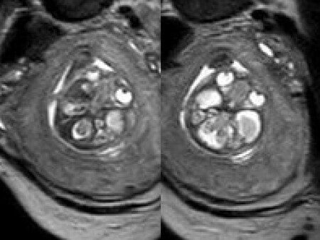
A 26-year-old primigravida with no significant past medical or family history was admitted at 18 weeks of gestation with preterm premature rupture of membranes. At 20 weeks of gestation, a detailed fetal ultrasound was performed under conditions of severe oligohydramnios, followed by fetal magnetic resonance imaging (MRI). Non-invasive prenatal testing (NIPT) for common aneuploidies was negative.


View the Answer Hide the Answer
Answer
We present a case of agenesis of the cavum septum pellucidum (CSP).
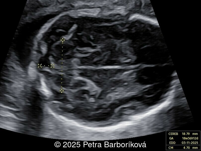
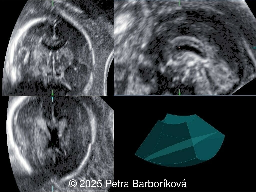
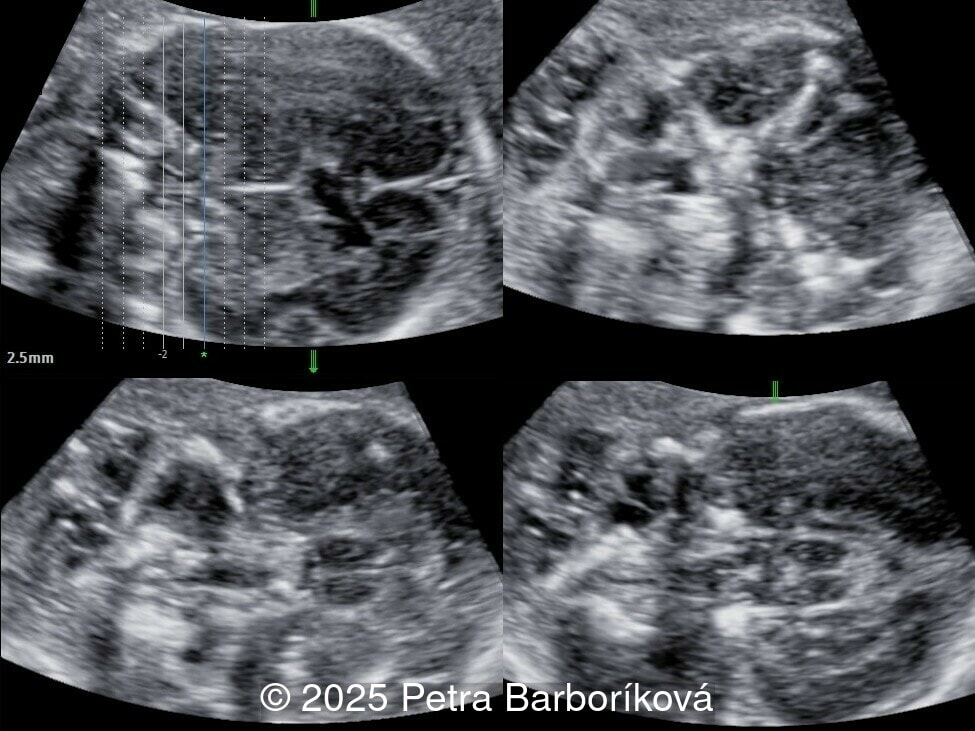
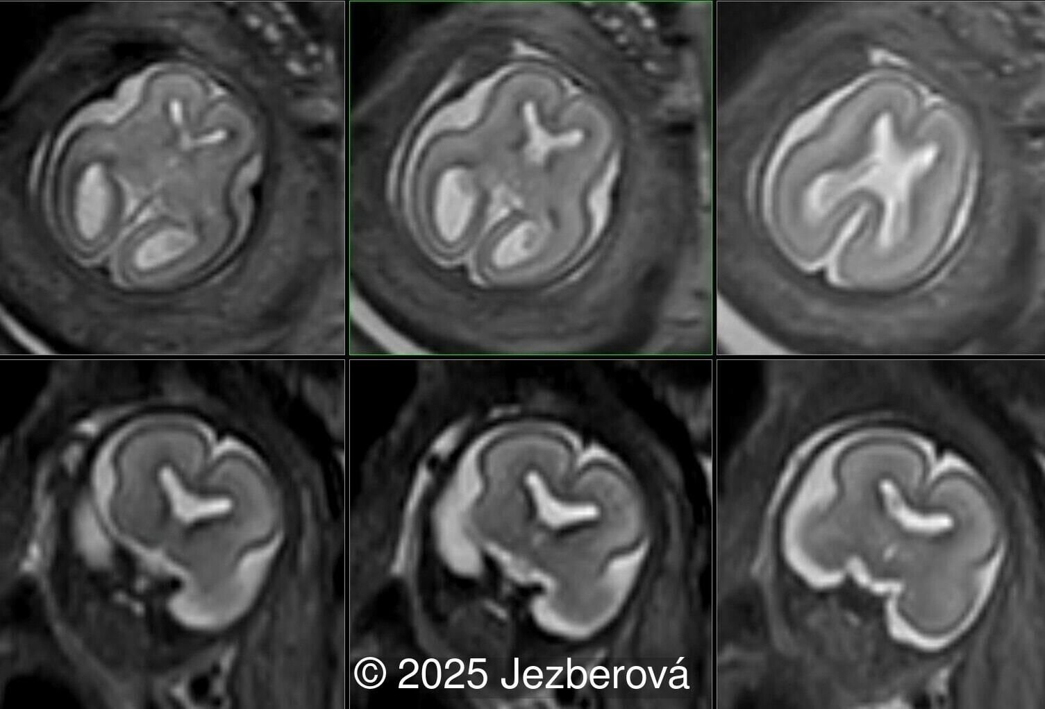
At 20 weeks, detailed ultrasound under conditions of severe oligohydramnios demonstrated absence of the typical box-shaped anechoic structure of the cavum septum pellucidum (CSP). The frontal horns of the lateral ventricles appeared narrow and pointed, giving the characteristic “bull’s horns sign.” Multiplanar and 3D reconstructions confirmed the presence of the corpus callosum, while the CSP was absent. Other midline and posterior fossa structures appeared normal. The optic chiasm was partially visualized, although assessment was limited by technical conditions. Fetal magnetic resonance imaging (MRI) was subsequently performed and confirmed isolated agenesis of the CSP with preserved corpus callosum and posterior fossa structures. The optic chiasm and pituitary stalk are visible in the sellar region, but the pituitary gland itself is not clearly identified in the sella turcica as it is a very small structure. No additional central nervous system anomalies were detected.
Following the diagnosis, the patient was referred for genetic counseling. Non-invasive prenatal testing was performed and was negative. Amniocentesis was offered but not performed due to the increased risk related to preterm premature rupture of membranes and the patient’s preference. During hospitalization, she received antibiotic therapy and antenatal corticosteroids as prevention for neonatal respiratory distress syndrome. At 26 weeks, a cesarean section was indicated due to fetal hypoxia. A female infant was delivered with extremely low birth weight, weighing 770g. Immediate postnatal management included intubation, surfactant administration, and prolonged ventilatory support.
The neonatal course was complicated by recurrent transfusions, systemic fungal infection requiring antifungal treatment, hyperbilirubinemia treated with phototherapy, bronchopulmonary dysplasia, and retinopathy of prematurity stage II. Postnatal cranial ultrasound confirmed the isolated agenesis of the CSP, with preserved corpus callosum and optic chiasm, and no additional malformations. During the hospitalization, the infant developed suspected central adrenal insufficiency (central hypoadrenocorticism), thought to be associated with agenesis of the CSP and hydrocortisone replacement therapy was initiated. Further endocrine evaluation found normal levels of adrenocorticotropic hormone, thyroid stimulating hormone, free thyroxine, growth hormone, and glycemia. Therefore, the low cortisol levels at birth may be related to transient immaturity rather than the CSP agenesis or septo-optic dysplasia.
At the time of discharge, the infant weighed 2000g and was feeding well on expressed breast milk and Neocate formula. Respiratory support was discontinued, with stable spontaneous ventilation. Neurological examination was adequate for corrected age, and psychomotor development was appropriate. She remains under multidisciplinary follow-up including endocrinology, neurology, ophthalmology, pulmonology, and pediatrics.
Discussion
The CSP is a transient developmental structure of the brain, representing a fluid-filled cavity between the two leaves of the septum pellucidum. Under physiological conditions, it can be visualized by ultrasound between 18 and 37 weeks of gestation [1-3]. Its absence during this period is considered pathological, though after 37 weeks of gestation, it begins to close spontaneously and is fused by three to six months of age [1,3-4].
Truly isolated agenesis of the CSP is extremely rare. In approximately 80-92% of cases, CSP agenesis is associated with major central nervous system malformations, most commonly schizencephaly, holoprosencephaly, severe ventriculomegaly, and agenesis of the corpus callosum [5,6]. In seemingly isolated agenesis of the CSP, this finding may be a clue to the diagnosis of a rare midline anomaly known as septo-optic dysplasia, which presents as a triad of absent CSP, optic nerve hypoplasia, and pituitary anomalies with endocrine impairment [6,7]. While only 40% of patients with septo-optic dysplasia have all three features, the clinical diagnosis is made when patients present with at least two of the abnormalities [8]. In our case, the optic chiasm was present, and the infant developed adrenal insufficiency after birth, though this was thought to be related to immaturity as further endocrine workup was normal. In studies reviewing fetuses with seemingly isolated CSP agenesis, 20-40% of cases were diagnosed with septo-optic dysplasia [6,7]. The pituitary gland [9] and optic chiasm [10] can be visualized in utero, however, the function can only be determined after birth [6]. Therefore, fetuses presenting with agenesis of the CSP, even if the optic chiasm and nerves appear normal on prenatal ultrasound, should be monitored for endocrine dysfunction postnatally.
The long-term neurodevelopmental outcome of isolated CSP agenesis has been reported as generally favorable [7,11]. The septum pellucidum is part of the limbic system, playing a role in emotional response, attention and activity, thus agenesis of this structure could lead to behavioral, learning and psychiatric disorders that may appear later in life [4]. A metanalysis of 46 infants with isolated CSP found that 6.5% were diagnosed with a major neurologic disability [7]. In another study, only 8% of school-aged children with isolated CSP on prenatal MRI required special teaching assistance in school [11]. However, patients with CSP agenesis and a diagnosis of septo-optic dysplasia have worse neurologic outcomes compared to their counterparts without septo-optic dysplasia [6], with 66% affected by developmental delay or seizures [8]. This makes prenatal counseling very difficult since the definitive diagnosis of septo-optic dysplasia cannot be made prenatally, and infants with this condition have worse neurodevelopmental outcomes than infants with isolated CSP agenesis.
This case illustrates the feasibility of prenatal diagnosis of agenesis of the CSP using ultrasound and fetal MRI, and emphasizes the importance of careful evaluation of the corpus callosum, optic pathways, and hypothalamic–pituitary axis. It is important to note that even morphologically isolated agenesis may be accompanied by endocrine abnormalities postnatally and a multidisciplinary team should evaluate for septo-optic dysplasia. In this case, the prognosis is determined not only by isolated CSP agenesis but also by complications of extreme prematurity.
References
[1] Timor-Tritsch IE, Monteagudo A, Pilu G. Ultrasonography of the Prenatal Brain. 4th ed. New York: McGraw Hill; 2023.
[2] Paladini D, Volpe P. Ultrasound of the fetal brain: normal development and cerebral pathologies. Cham: Springer; 2022.
[3] Falco P, Gabrielli S, Visentin A, et al. Transabdominal sonography of the cavum septum pellucidum in normal fetuses in the second and third trimesters of pregnancy. Ultrasound Obstet Gynecol. 2000 Nov;16(6):549-53.
[4] Siala S, Homen D, Smith B, et al. Imaging of the septum pellucidum: normal, variants and pathology. Br J Radiol. 2023 May 24;96(1151):20221058.
[5] Belhocine O, André C, Kalifa G, et al. Does asymptomatic septal agenesis exist? A review of 34 cases. Pediatr Radiol. 2005 Apr;35(4):410-8.
[6] Shinar S, Blaser S, Chitayat D, et al. Long-term postnatal outcome of fetuses with prenatally suspected septo-optic dysplasia. Ultrasound Obstet Gynecol. 2020 Sep;56(3):371-377.
[7] Di Pasquo E, M Kuleva M, Arthuis C, et al. Prenatal diagnosis and outcome of fetuses with isolated agenesis of septum pellucidum: systematic review and meta-analysis. Ultrasound Obstet Gynecol. 2022 Feb;59(2):153-161.
[8] Nagasaki K, Kubota T, Kobayashi H, et al. Clinical characteristics of septo-optic dysplasia accompanied by congenital central hypothyroidism in Japan. Clin Pediatr Endocrinol. 2017;26(4):207-213.
[9] Katorza E, Bault JP, Gilboa Y, et al. Prenatal visualization of the pituitary gland using 2- and 3-dimensional sonography: comparison to prenatal magnetic resonance imaging. J Ultrasound Med. 2012 Oct;31(10):1675-80.
[10] Alonso I, Azumendi G, Romero M, et al. Fetal optic chiasm: three steps for visualization and measurement on routine transabdominal ultrasound. Ultrasound Obstet Gynecol. 2019 Jul;54(1):135-136.
[11] Righini A, Izzo G, Doneda C, et al. School-Age Outcome of Fetuses with Isolated Complete Septum Pellucidum Agenesis at Prenatal Magnetic Resonance Imaging. Neuropediatrics. 2022 Feb;53(1):26-31.
Discussion Board
Winners
Javier Cortejoso Spain Physician
paola quaresima Italy Physician

belen garrido Spain Physician
Mayank Chowdhury India Physician
Shilpen Gondalia India Physician
carlos lopez Venezuela Physician

CHEN YANG China Physician
Victoria Giang Viet Nam Physician
Amparo Gimeno Spain Physician
Elena Andreeva Russian Federation Physician
ALBANA CEREKJA Italy Physician
Eti Zetounie Israel Sonographer
Murat Cagan Turkey Physician
ANA PAULA PASSOS Brazil Physician
Ionut Valcea Romania Physician
Đặng Mai Quỳnh Viet Nam Physician
Hien Nguyen Van Viet Nam Physician
Almaz Kinzyabulatov Russian Federation Physician
Malek Nassar Lebanon Physician
Walter Plasencia Spain Physician
Zuzana Briešková Slovakia Physician
Martina Vagaská Slovakia Physician

Thomson Thomson Indonesia OB-GYN
Annette Reuss Germany Physician
Arati Appinabhavi India Physician

YULIA VISHNEVSKAYA Russian Federation Physician
shay kevorkian Israel Physician
Costin Radu Lucian Romania Physician
Dr Dhara patel India Physician
Hana Habanova Slovakia Physician
Marius Bogdan Muresan Romania Physician
Leslie Bowden United States Sonographer
ASHLEA HARDIN United States Sonographer
Ali Ozgur Ersoy Turkey Physician
Ali Karaege Turkey Physician
everardo GUANABARA Brazil Physician
Albert Guarque Rus Spain Physician
jimena salcedo Spain Physician
Luu Anh Viet Nam Physician
ANDRES ARENCIBIA MOLINA United States Physician