Case of the Week #618
(1) Kenyatta National Hospital, Kenya; (2) UCSF Health, San Francisco, CA, United States
34-year-old primigravida was referred at 32 weeks due to a routine ultrasound indicating a cardiac mass at 30 weeks.


View the Answer Hide the Answer
Answer
We present a case of Cardiac rhabdomyoma associated with Tuberous Sclerosis.
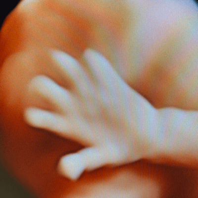
Our images demonstrated a large, homogenous echogenic mass on the wall of the left ventricle and apex without significant outflow tract obstruction (Image 2, 3, Video 1). Images of the fetal brain showed multiple echogenic foci (Image 4, 5).
The fetus had a normal cardiac function until term. At 37 weeks, an elective Caesarean section was performed and the neonate delivered with APGAR scores of 7, 8, 8 at 1, 5, and 10 minutes. An echocardiogram at birth revealed multiple masses on the myocardium involving the left and right ventricular walls, likely rhabdomyoma, a small patent ductus arteriosus, mild persistent pulmonary hypertension of the newborn and a pericardial effusion. The neonate had intact cardiac septa and normal pulmonary and systematic venous return. On day 6 of life, the neonate was started on sildenafil 2mg/kg due to persistently low saturations and oxygen requirement. A repeat ECHO performed one week after delivery revealed normal cardiac function, myocardium masses and minimal pericardial effusion. An MRI of the brain at four weeks of life and revealed cortical lesions, and subependymal hamartomas bilaterally. These features, cortical tubers and cardiac rhabdomyoma, were most consistent with tuberous sclerosis. No genetic testing was performed due to poor maternal resources.
Discussion
Fetal cardiac tumors are rare with an incidence of approximately 0.03-0.14% [1,2]. Rhabdomyoma is the most common cardiac tumor in fetal life, encompassing approximately 60% of fetal cardiac tumors [3]. In a metanalysis reviewing 138 cases of fetal rhabdomyoma, diagnosis occurred more commonly after 24 weeks gestation and nearly 90% of the tumors were located in the ventricles or interventricular septum [4]. Few cases presented with congenital heart defects (4%), chromosomal abnormalities (2%), and extracardiac anomalies (1.4%) [4]. Larger tumors are more likely to can cause hemodynamic disturbance and dysrhythmia. Therefore, poor prognostic indicators include tumor size >20mm, fetal dysrhythmia, and fetal hydrops [4]. Perinatal death occurs in 13% of cases [5].
The differential diagnosis for a cardiac mass includes fibroma, myxoma, teratoma, and hemangioma. On prenatal ultrasound, rhabdomyomas appear as round, homogeneous masses in the ventricles. Fibromas are hyperechogenic tumors associated with calcifications and cystic degenerations. Teratomas are located in the pericardial cavity and associated with pericardial effusion. Hemangiomas are usually located in the right atrium and show more complex echogenicity with both solid and cystic portions mixed with calcifications [4].
Tuberous sclerosis is an autosomal dominant condition first described by von Recklinhausen in 1862 and caused by a mutation in either the TSC1 gene, coding for hamartin, or the TSC2 gene, coding for tuberin [6]. Hamartin and tuberin are components of the TSC complex which regulates cell growth and proliferation by inhibiting the mammalian target of rapamycin (mTOR) signaling pathway. Mutations in these components results in inactivation of the TSC complex and upregulation of mTOR which leads to the formation of benign hamartomas in the body [6]. The incidence of tuberous sclerosis is 1 in 6,760 to 13,520 live births [7]. Clinical diagnosis requires two major features, which include skin, brain, heart, retina, lung and kidney lesions, or the combination of one major feature and two minor features [8]. Patients with tuberous sclerosis are affected with epilepsy in approximately 70-80% of cases, as well as neurodevelopmental dysfunction [7,9].
In the prenatal period, patients with tuberous sclerosis may present with cardiac rhabdomyoma, cortical tubers, subependymal nodules, renal cysts, and rarely, subependymal giant cell astrocytoma [6]. Neurologic lesions are usually detected late second trimester or third trimester of pregnancy. If no extracardiac manifestations are identified, multiple cardiac rhabdomyoma has been associated with tuberous sclerosis in utero [6]. A metanalysis found that the overall incidence of tuberous sclerosis was 64% in fetuses with cardiac rhabdomyoma, and a family history of tuberous sclerosis or multiple cardiac tumors was associated with the presence of tuberous sclerosis [4]. In a study reviewing 240 cases of fetal cardiac rhabdomyoma, 66% of cases had multiple cardiac tumors, 41% had cortical tubers or subependymal nodules detected an average of four weeks after the cardiac rhabdomyoma, and 65% had a genetic diagnosis of tuberous sclerosis. Additional analysis showed that if fetuses present with multiple cardiac tumors, a genetic mutation causing tuberous sclerosis was found in 75% [6].
Cardiac rhabdomyoma often regress spontaneously with good prognosis. Surgical resection may be necessary in cases with large tumors causing intracardiac obstruction, though may be difficult with multifocal tumors or in preterm infants [10]. Everolimus is a serine-threonine kinase mTOR inhibitor which leads to alteration of cell protein synthesis and prevention of cell proliferation, differentiation, growth and migration. In neonates, with cardiac rhabdomyoma, treatment with Everolimus results in the regression of the cardiac tumor approximately 12 times faster in the historical controls [10]. Similarly, maternal administration of mTOR inhibitors has been shown to be effective in treating hemodynamically significant fetal cardiac rhabdomyomas [11].
References
[1] Holley DG, Martin GR, Brenner JI, et al. Diagnosis and management of fetal cardiac tumors: a multicenter experience and review of published reports. J Am Coll Cardiol. 1995 Aug;26(2):516-20.
[2] Okmen F, Ekici H, Hortu I, et al. Outcomes of antenatally diagnosed fetal cardiac tumors: a 10-year experience at a single tertiary referral center. J Matern Fetal Neonatal Med. 2022 Sep;35(18):3489-3494.
[3] Isaacs H. Fetal and Neonatal Cardiac Tumors. Pediatr Cardiol. 2004 May-Jun;25(3):252-73.
[4] Chao AS, Chao A, Wang TH, et al. Outcome of antenatally diagnosed cardiac rhabdomyoma: case series and a meta-analysis. Ultrasound Obstet Gynecol. 2008 Mar;31(3):289-95.
[5] Yinon Y, Chitayat D, Blaser S, et al. Fetal cardiac tumors: a single-center experience of 40 cases. Prenat Diagn. 2010 Oct;30(10):941-9.
[6] Milon V, Malinge MC, Blanluet M, et al. Diagnosis of tuberous sclerosis in the prenatal period: a retrospective study of 240 cases and review of the literature. Eur J Hum Genet. 2024 May 28.
[7] Ebrahimi-Fakhari D, Mann LL, Poryo M, et al. Incidence of tuberous sclerosis and age at first diagnosis: new data and emerging trends from a national, prospective surveillance study. Orphanet J Rare Dis. 2018 Jul 17;13(1):117.
[8] Northrup H, Aronow ME, Martina Bebin E, et al. Updated International Tuberous Sclerosis Complex Diagnostic Criteria and Surveillance and Management Recommendations. Pediatr Neurol. 2021 Oct:123:50-66.
[9] Peng L Cai Y, Wu J, et al. Prenatal diagnosis and clinical management of cardiac rhabdomyoma: a single-center study. Front Cardiovasc Med. 2024 Feb 16;11:1340271.
[10] Aw F, Goyer I Raboisson MJ, et al. Accelerated Cardiac Rhabdomyoma Regression with Everolimus in Infants with Tuberous Sclerosis Complex. Pediatr Cardiol. 2017 Feb;38(2):394-400.
[11] Ebrahimi-Fakhari D, Stires G, Hahn E, et al. Prenatal Sirolimus Treatment for Rhabdomyomas in Tuberous Sclerosis. Pediatr Neurol. 2021 Dec:125:26-31.
Discussion Board
Winners

Dianna Heidinger United States Sonographer
Javier Cortejoso Spain Physician
paola quaresima Italy Physician

Pawel Swietlicki Poland Physician
Igor Yarchuk United States Sonographer
Chursina Olga Russian Federation Physician
belen garrido Spain Physician
Andrii Averianov Ukraine Physician

Ana Ferrero Spain Physician
Mayank Chowdhury India Physician
Aizirek Ganieva Kyrgyzstan Physician
RITIKA BHANDARI India Physician
Vladimir Lemaire United States Physician
Ivan Ivanov Russian Federation Physician
Boujemaa Oueslati Tunisia Physician

Tatiana Koipish Belarus Physician
CHARLES SARGOUNAME India Physician
Aysegul Ozel Turkey Physician
Irvin Jacob Vélez Machorro Mexico Physician
Alvaro Gómez Mexico Physician
Kimberly Delaney United States Sonographer
Olivia Ionescu United Kingdom Physician
Marianovella Narcisi Italy Physician
Tudor Iacovache Romania Physician
Annette Reuss Germany Physician
Amparo Gimeno Spain Physician
Elena Andreeva Russian Federation Physician
CRISTINA MARTINEZ PAYO Spain Physician
ALBANA CEREKJA Italy Physician
Eti Zetounie Israel Sonographer
Eda Özden Tokalıoğlu Turkey Physician
Deval Shah India Physician
SAMUEL GELVEZ TELLEZ Colombia Physician
Murat Cagan Turkey Physician

Sonio Sonio France AI
Charlotte Conturie United States Physician
Loai Said Palestinian Territory, Occupied Physician
gholamreza azizi Iran, Islamic Republic of Physician
Fatih Akkuş Turkey Physician
Gayane Begjanyan Armenia Physician
Ionut Valcea Romania Physician
Viktoriya Shuman United States
aastha mehra India Physician
Đặng Mai Quỳnh Viet Nam Physician
Hien Nguyen Van Viet Nam Physician

Viralkumar Madhu India Sonographer
Borisova Elena Russian Federation Physician
Miguel Sanchez Mexico Physician
Almaz Kinzyabulatov Russian Federation Physician
Kareem Haloub Australia Physician
Vitor Oliveira Brazil Physician

Pradeep Kumar India Physician
Zuzana Briešková Slovakia Physician

abdullah sarıyıldırım Turkey Physician
Fred Pop Uganda Sonographer
shruti Agarwal India Physician

CHERYL TURNER United States Sonographer
Brindusa Ciobanu Romania Physician
shay kevorkian Israel Physician
Sruthi Pydi India Physician
Laura Wharton United Kingdom Physician
zozo sichala Zambia radiology technologist
Shina Kaur India Physician
Deniz Delibaş Turkey Physician
Rupal Sasani India Physician
Hiral Shah India Fetal Medicine specialist
Nestor Ferrer Venezuela Physician
philip pattyn Belgium Physician
Anastasia Igolkina Russian Federation Physician
Nikhila BL India Physician
Eylem Eşsizoğlu Turkey Physician
Anh Duy Nguyen Viet Nam Sonographer
Petra Barboríková Slovakia Physician
Dolly Agrawal India Physician
Magdalena Piekarska Poland Physician
Nguyễn Lê Hoàng Viet Nam Physician
Denys Saitarly Israel Physician
Le Tien Dung Viet Nam Physician
Tetiana Ishchenko Ukraine Physician
Costin Radu Lucian Romania Physician
Almudena Martín García Spain Physician
María Victoria Peral Parrado Spain Physician
Hetal Patel India Physician
Liliya Lazukina Russian Federation Physician
Le Duc Viet Nam Physician
Mậu Nguyễn Bá Viet Nam Physician

Dr Dhara patel India Physician
Hana Habanova Slovakia Physician
Yücel Kaya Turkey Physician
Joanne Maloney United States Sonographer

Dr Mayur C Trivedi India Physician
Gnanasekar Periyasamy India Physician
Achmad Feryanto Indonesia Physician
Umesh Athavale India Physician
Navya Sri Mopada India Physician
Jagdish Suthar India Physician
Stefan Schmitt- Heidsieck Germany Physician
Dr Othman Alasali Jordan Physician
CARLOS JOSE PIÑA VILLEGAS Peru Physician
Uyên Huỳnh Viet Nam Physician
Akhani Madhvi India Physician

Fco Javier Martínez Cortés Spain Physician
Gabriel Bogdan France Physician
Viktoriia Putsenko Russian Federation Physician
Ramya Smruthi DHATRIC India Physician
Uğur Yıldırım Turkey Physician
Harshini Battula India Physician

Dr. Amol Deshmukh India Physician
Yugandhara Kamble India Physician
kristel villanueva Venezuela Physician
Gaea Moore United States Physician