Case of the Week #611
Hospital Universitario de Canarias; Hospital Universitario Doctor José Molina Orosa
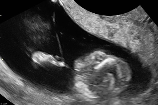
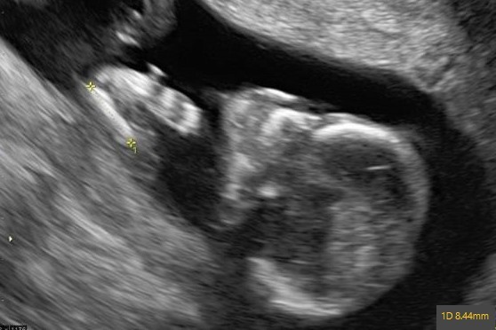
A 30-year-old primigravida presents for a routine 1st trimester scan at 13 weeks gestation. Her past medical and family history are unremarkable. These are some of the images we obtained.

View the Answer Hide the Answer
Answer
We present a case of Amniotic Band Sequence (ABS) that led to a defect at the level of the left upper limb.
Our images reveal a small radius, ulna and left hand with radial deviation. We can observe the presence of a narrowing at the level of the wrist, with visualization of a membrane at this level suggesting an amniotic band.
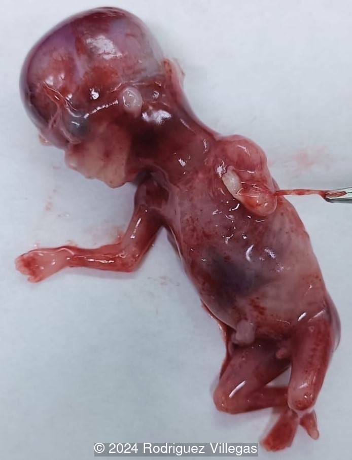
After the diagnosis, the patient declined invasive testing and terminated the pregnancy. The genetic test (CGH-array) was normal (46 XY), and the fetal autopsy revealed hypoplasia of the left upper limb in all its parts (type V according to Swanson's classification) [1].
Discussion
Amniotic bands are caused early in embryo development and lead to fetal amputations or constrictions. As a consequence of its movements, the fetus is at risk of entanglement in these bands, leading to malformations. It occurs sporadically with an incidence between 1 in 8600 [1] to 1 in 10,000 live births [2], though rare familial cases have been reported [2]. Amniotic band attachment can be categorized into constrictive bands, limb anomalies, craniofacial anomalies and neural tube-like anomalies [3]. Limb deformities are the most frequent manifestation with the upper limbs more commonly affected [2]. The constriction of a normally developing structure by an amniotic band can be confined to the skin and soft tissue causing a constriction band, or they may compromise the vascular supply, lymphatic system, bone, and nerves, resulting in lymphedema, neuropathy, fracture, and amputation. If it happens early in pregnancy, the devitalized body part is resorbed over time. Adherence of the amniotic band, even without constriction, can have adverse mechanical effects that result in malformation or deformation [4]. Anomalies of the skull, face, and body wall can be seen and include cleft lip and palate, asymmetric microphthalmos, nasal deformities, asymmetric cephalocele, anencephaly, acrania, gastroschisis, omphalocele, and bladder exstrophy [4,5].
Prenatally, the diagnosis is suspected by ultrasound detection of constriction rings, distal limb edema, limb amputations, or lateralization of body wall or craniofacial abnormalities. The majority of defects seen with amniotic band syndrome are asymmetric, thus lateralization of a usually midline disease process, such as cephaloceles [4], should raise suspicion for amniotic bands [3]. In some cases, thin strands of amnion can be seen crossing the gestational sac and adherent to the fetus, restricting its movement. Manipulation of the maternal position to reveal a tethering band can be helpful in making the diagnosis [3].
The pathophysiology is not well understood and both an intrinsic and extrinsic theory have been proposed: the intrinsic theory describes an early embryonic defect, while the extrinsic theory suggests rupture of the amniotic sac, allowing the fetus to enter the chorionic cavity and attach to fibrous mesodermal bands [1,3]. The extrinsic theory does not easily explain anomalies such as imperforate anus, tracheoesophageal fistula, and septo-optic dysplasia [5]. Additionally, a vascular theory postulates that amniotic bands and certain congenital defects have a common pathway related to a vascular insult. Congenital vascular anomalies have been found in patients with amniotic band sequence [6]. Furthermore, conditions associated with relative hypoxia, such as smoking and living at altitude, have been associated with amniotic bands [1]. Other risk factors for amniotic band sequence may include smoking, drug use, amniocentesis or uterine surgery, and twin pregnancy [1].
Amniotic band sequence has a wide clinical spectrum; from a single mild abnormality with an excellent prognosis to multiple severe anomalies affecting the cranium, umbilical cord, and body wall that are incompatible with life [7-9]. Fetoscopic procedures have been attempted to salvage limbs. The ideal time for fetoscopic intervention is before vascular occlusion occurs when arterial waveforms are abnormal, but flow is patent and extremity edema is present [10].
References
[1] Barzilay E, Harel Y, Haas J, et al. Prenatal diagnosis of amniotic band syndrome - risk factors and ultrasonic signs. J Matern Fetal Neonatal Med. 2015 Feb;28(3):281-3
[2] Lowry RB, Bedard T, Sibbald B. The prevalence of amnion rupture sequence, limb body wall defects and body wall defects in Alberta 1980-2012 with a review of risk factors and familial cases. Am J Med Genet A. 2017 Feb;173(2):299-308.
[3] Jensen KK, Oh KY, Kennedy AM, et al. Intrauterine Linear Echogenicities in the Gravid Uterus: What Radiologists Should Know. Radiographics. 2018 Mar-Apr;38(2):642-657.
[4] Weinstein B, Hassouba M, Flores, RL, et al. Digital-Facial Translocation in Amniotic Band Sequence: Evidence of the Intrinsic Theory. J Craniofac Surg. 2018 Oct;29(7):1890-1892.
[5] Gonçalves LF, Jeanty P. "Amniotic band syndrome." TheFetus.net. https://thefetus.net/content/amniotic-band-syndrome-4/, Publish date 5/2002.
[6] Daya M, Makakole M. Congenital vascular anomalies in amniotic band syndrome of the limbs. J Pediatr Surg. 2011 Mar;46(3):507-13.
[7] Chen CP, Chang TY, Lin YH, et al. Prenatal sonographic diagnosis of acrania associated with amniotic bands. J Clin Ultrasound. 2004 Jun;32(5):256-60
[8] Lurie S, Feinstein M, Mamet Y. Umbilical cord strangulation by an amniotic band resulting in a stillbirth. J Obstet Gynaecol Res. 2008 Apr;34(2):255-7.
[9] Chen CP. Prenatal diagnosis of limb-body wall complex with craniofacial defects, amniotic bands, adhesions and upper limb deficiency. Prenat Diagn. 2001 May;21(5):418-9.
[10] Javadian P, Shamshirsaz AA, Haeri S, et al. Perinatal outcome after fetoscopic release of amniotic bands: a single-center experience and review of the literature. Ultrasound Obstet Gynecol. 2013 Oct;42(4):449-55.
Discussion Board
Winners

Dianna Heidinger United States Sonographer

Javier Cortejoso Spain Physician

Padman KG United Kingdom Sonographer
Pawel Swietlicki Poland Physician

Andrii Averianov Ukraine Physician

Ana Ferrero Spain Physician

Alexandr Krasnov Ukraine Physician

Mayank Chowdhury India Physician
Vera Osadshaya Russian Federation Physician

Vladimir Lemaire United States Physician

Tatiana Koipish Belarus Physician

Caroline Reichert Garcia Brazil Physician
Olivia Ionescu United Kingdom Physician
Marianovella Narcisi Italy Physician
Annette Reuss Germany Physician
Eti Zetounie Israel Sonographer

Deval Shah India Physician
Murat Cagan Turkey Physician
Moftah Almgrhi United States
gholamreza azizi Iran, Islamic Republic of Physician
Ionut Valcea Romania Physician

Sviatlana Akhramovich Belarus Physician
reyhan ayaz Turkey Physician
Borisova Elena Russian Federation Physician
Almaz Kinzyabulatov Russian Federation Physician

Kareem Haloub Australia Physician

abdullah sarıyıldırım Turkey Physician
shruti Agarwal India Physician
Lucy Bayer-Zwirello United States Physician
SAVITA SHIRODKAR India Physician

Lynn Davis United States Sonographer
Mary Jones United States Sonographer

Ismail Guzelmansur Turkey Physician
zozo sichala Zambia radiology technologist
Jacqueline L J Cullen United States Sonographer
Ashraf Elkashef Egypt Physician

philip pattyn Belgium Physician

Nguyễn Lê Hoàng Viet Nam Physician

Le Tien Dung Viet Nam Physician

Tetiana Ishchenko Ukraine Physician
Eve Palomino United States Sonographer
Nora Fox United States Sonographer

Hana Habanova Slovakia Physician

Tamara Yarygina Russian Federation Physician

Dr Mayur C Trivedi India Physician
Elena Kogteva Russian Federation Physician
Syeda Amna Mehmood United Arab Emirates Physician
Achmad Feryanto Indonesia Physician

Văn Kiệt Võ Viet Nam Physician

Jagdish Suthar India Physician
Rinku Vasaya Congo, The Democratic Republic of The Sonographer
Svetlana Bakhtiyarov United States Sonographer
Sonia Graham United States Sonographer