Case of the Week #606
Maternal Fetal Associates in Reston, Virginia
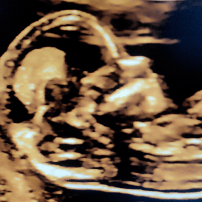
40-year-old multipara with type II diabetes presents at 16 weeks for genetic scan because she missed her 1st trimester screening exam. Ultrasound revealed the following findings. There were other numerous abnormalities in the rest of the scan apart from the ones shown here.


View the Answer Hide the Answer
Answer
We present a case of Trisomy 21. Non-invasive prenatal testing was concerning for Down's syndrome and amniocentesis confirmed the diagnosis.
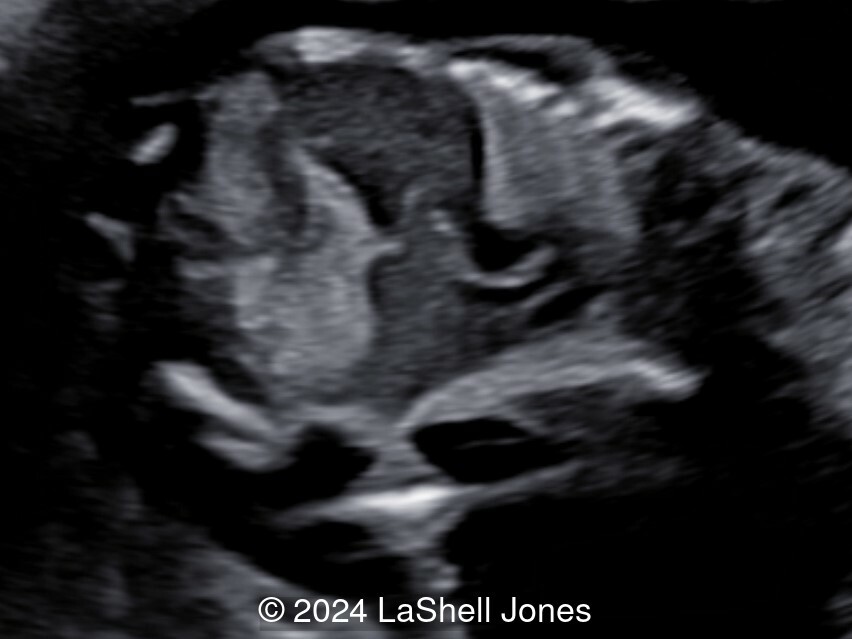
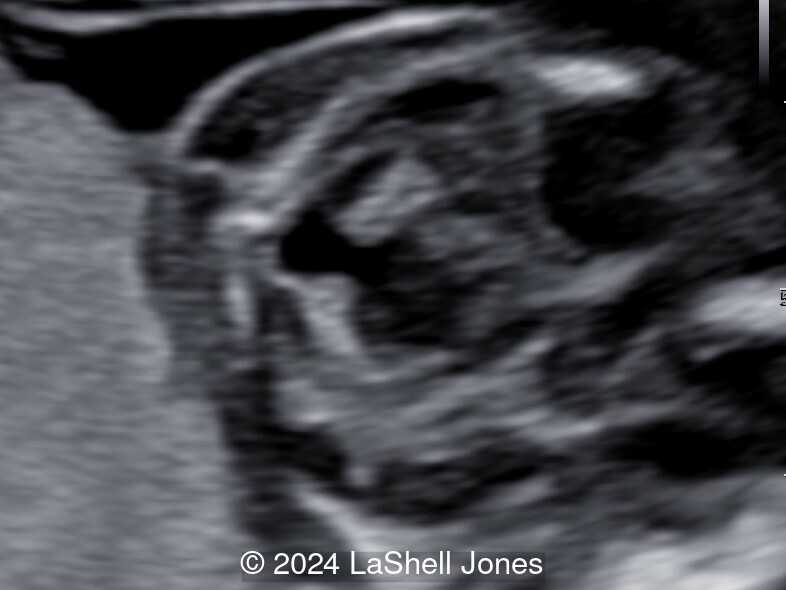
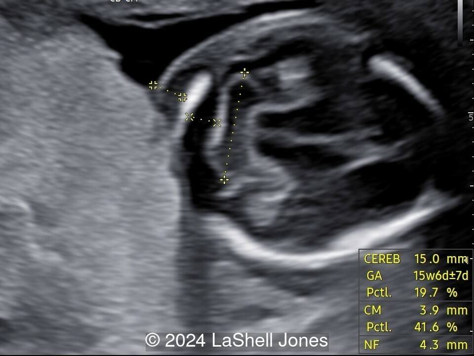
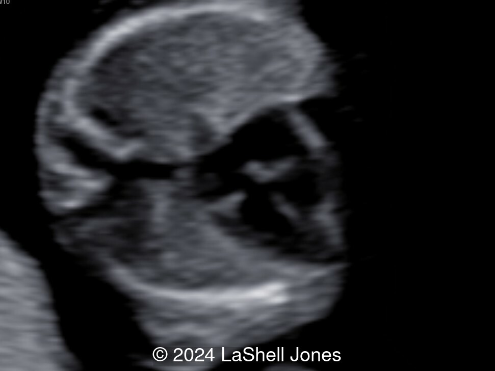
Our images demonstrated the following:
- Image 1: Absent Nasal Bone
- Image 2: liver calcifications
- Image 3-4: Echogenic bowel
- Image 5: "Keyhole" Dandy Walker Variant
- Image 6: Lagging Cerebellum measurement
- Image 7: VSD
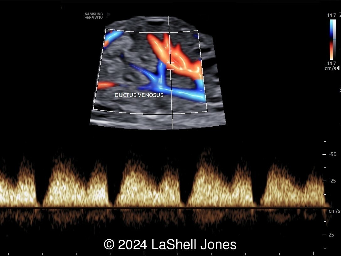
- Image 8: Absent A - wave Ductus Venosus
- Image 9: Pericardial Effusion
- Image 10: Thick Nuchal Fold
Discussion
Down Syndrome, also known as trisomy 21, is the most prevalent chromosomal abnormality observed in newborns. Approximately 6,000 infants are born with Down Syndrome annually in the United States, equating to a frequency of about 1 in every 700 births. Various prenatal genetic screening methods and diagnostic tests are utilized to accurately identify Down Syndrome and other aneuploidies during pregnancy.
Ultrasound plays a crucial role in the screening for aneuploidies. It enables the detection of both major structural abnormalities and minor soft markers in fetuses affected by aneuploidies. Down Syndrome can manifest with anomalies in various systems such as cardiovascular, central nervous, craniofacial, musculoskeletal, gastrointestinal, and urinary tract. Notably, major structural anomalies like duodenal atresia and cardiac defects such as septal defects, tetralogy of Fallot, and atrioventricular canal defects may not always be identified through prenatal ultrasound screening.
Nuchal translucency (NT) measurement in the first trimester is indicative of the subcutaneous fluid-filled space at the back of the fetal neck. An increased NT measurement is associated with a higher risk of aneuploidies, including Down Syndrome, with a detection rate ranging from 64% to 70%. Additionally, the presence of a nuchal cystic hygroma or a hypoplastic/absent nasal bone in the first trimester can also indicate a risk of aneuploidies, including Down Syndrome.
During the second trimester, soft markers such as echogenic intracardiac foci, pyelectasis, short femur length, choroid plexus cysts, echogenic bowel, thickened nuchal skin fold, and ventriculomegaly are commonly assessed. Some of these markers, like echogenic bowel, thickened nuchal fold, and ventriculomegaly, have high likelihood ratios, prompting further genetic counseling and additional screening or diagnostic tests. Conversely, markers with lower predictive values may also warrant aneuploidy screening if not previously conducted.
While ultrasound is a valuable tool in identifying potential markers for Down Syndrome, it should not be solely relied upon for diagnosis. Combining ultrasound findings with maternal serum screening tests, such as first and second trimester screening, as well as integrated and sequential screening, enhances the sensitivity for detecting Down Syndrome. Cell-free DNA testing boasts a high detection rate of 99% for Down Syndrome. Diagnostic procedures like amniocentesis or chorionic villus sampling should be considered when screening results indicate a positive likelihood of aneuploidies.
References
American College of Obstetricians and Gynecologists’ Committee on Practice Bulletins—Obstetrics; Committee on Genetics; Society for Maternal-Fetal Medicine. Screening for Fetal Chromosomal Abnormalities: ACOG Practice Bulletin, Number 226. Obstet Gynecol. 2020 Oct;136(4):e48-e69.
Discussion Board
Winners

Dianna Heidinger United States Sonographer

Javier Cortejoso Spain Physician

Padman KG United Kingdom Sonographer

Igor Yarchuk United States Sonographer

Ana Ferrero Spain Physician

Vladimir Lemaire United States Physician

Tatiana Koipish Belarus Physician

CHARLES SARGOUNAME India Physician
Aysegul Ozel Turkey Physician

ALBANA CEREKJA Italy Physician

Deval Shah India Physician
Murat Cagan Turkey Physician
Mesud Sehic Bosnia and Herzegovina Physician
gholamreza azizi Iran, Islamic Republic of Physician
Gayane Begjanyan Armenia Physician
Ionut Valcea Romania Physician
reyhan ayaz Turkey Physician

Kathrine Montagne United States Sonographer
Zuzana Briešková Slovakia Physician

CHERYL TURNER United States Sonographer
Perrine Riou-Kerangal French Polynesia Sage-femme échographiste

Veronika Bartkovjaková Slovakia Physician
Petra Barboríková Slovakia Physician

Nguyễn Lê Hoàng Viet Nam Physician

Denys Saitarly Israel Physician

Le Tien Dung Viet Nam Physician
Costin Radu Lucian Romania Physician
Gaurav Sharma India Physician

Le Duc Viet Nam Physician
Erika Zanzarelli Italy Physician

Aigerim Nukenova Kazakhstan Physician
Rebekah Matherly United States Sonographer
PADMA Priya DHARSHINI India Physician

Hana Habanova Slovakia Physician
Anjali Gupta India Physician
Mithun Chowdhury Bangladesh Physician
ELHAM M Saudi Arabia Physician
MARIA JUSTINA AGUERRE GOGORZA Spain Physician
Syeda Amna Mehmood United Arab Emirates Physician
Mia Noser Switzerland Physician
Luan Nguyen Thanh Viet Nam Physician
Quan Le Viet Nam Physician
Angel Barco United Arab Emirates Sonographer