Case of the Week #588
Miras, Medical clinic, Sterlitamak, Republic of Bashkortostan, Russian Federation
A 28-year-old G2P1 woman presented to our clinic at 32 weeks, 6 days gestation for routine third trimester ultrasound screening. The following images were obtained.
View the Answer Hide the Answer
Answer
We present a case of cervical cancer during the pregnancy. The patient presented with genital bleeding and a cervical mass suggestive of malignancy at 24 weeks of pregnancy. Diagnosis of cervical cancer (FIGO IIB) was made by Pap smear, cervical biopsy and MRI. In addition, DNA test detected the presence of the human papillomavirus (HPV 16 and 18). Ultrasound examination did not reveal any fetal abnormalities besides polyhydramnios (AFI 29.3 cm). During the transvaginal cervical assessment, we observed the following ultrasound changes of the cervix, which are common for cervical cancer.
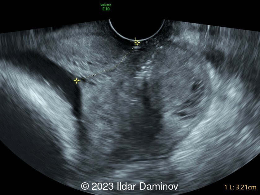
- Image 1, video 1 shows a large irregular mass invading mainly the posterior wall of the uterine cervix and containing cystic spaces.
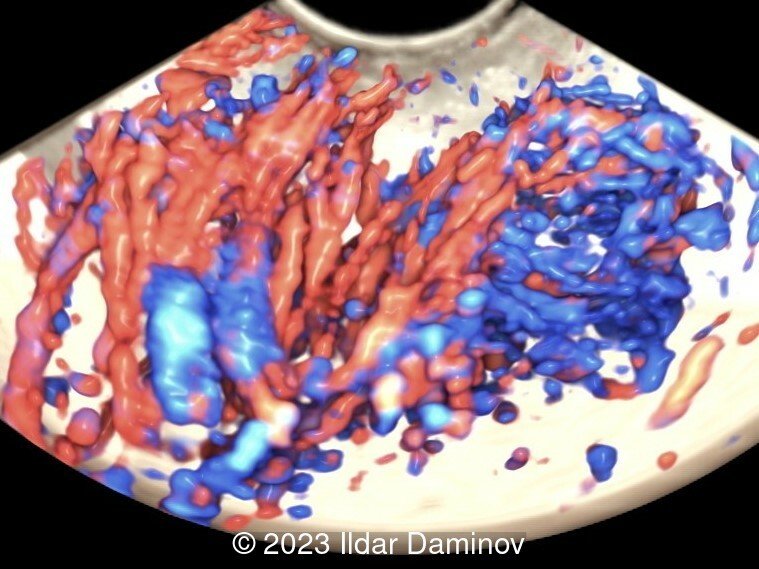
- Image 2, video 2 demonstrates an abundant vascularization of the cervix due to tumor angiogenesis.
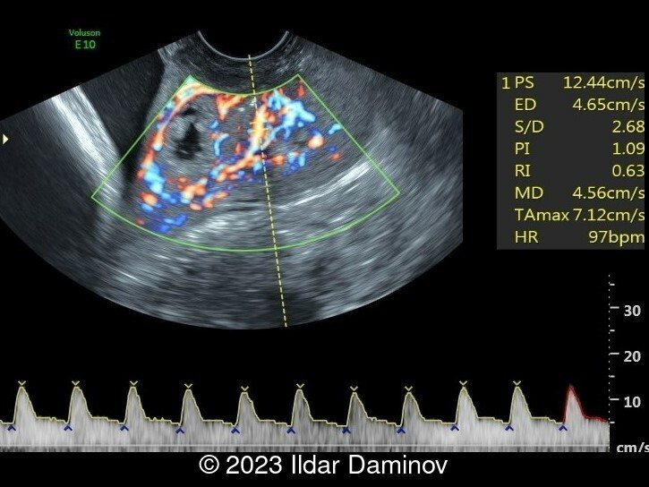
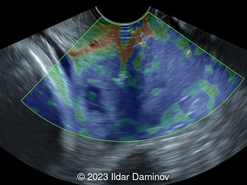
- Images 3 and 4 show intratumoral blood flow measurement and assessment of cervical stiffness by elastography. A blue coloring prevails, which is more common in malignant tumors.
Emergency cesarean section was performed at 34 weeks of gestation due to premature rupture of amniotic membranes, and a male newborn was delivered (weight 2000g, height 45cm and Apgar score 5/7). The postoperative period was uneventful, and the patient was discharged on 7th postoperative day in good health. Postnatal adaptation of the newborn was without complications. Further management of cervical cancer involved chemoradiotherapy treatments. Despite therapy, the woman died six months later.
Discussion
Cervical cancer during pregnancy is relatively uncommon. Incidence rates vary from 0.1 to 12 per 10,000 pregnancies [1,2]. Early cervical cancer diagnosis is based on results derived from the Pap smear test, colposcopy and diagnostic biopsies. Cervical cancer is clinically staged according to FIGO guidelines, though there is increased use of cross sectional imaging modalities (CT, MRI, PET-CT) to evaluate important prognostic factors such as tumor size, parametrial invasion, endocervical extension, pelvic side wall or adjacent/distal organ involvement, and lymph node status [3,4]. The treatment of cervical cancer in pregnancy is complex as both the optimal oncologic therapy as well as the preservation of the health of the fetus should be considered. Treatment options include conservative and surgical approaches based on tumor size, lymph node involvement, gestational age and the patient’s wish to continue the pregnancy [5,6].
References
[1] Han SN, Gziri MM, Van Calsteren K, et al. Cervical cancer in pregnant women: treat, wait or interrupt? Assessment of current clinical guidelines, innovations and controversies. Ther Adv Med Oncol. 2013 Jul; 5(4):211-9.
[2] Al-Halal H, Kezouh A, Abenhaim H. Incidence and obstetrical outcomes of cervical intraepithelial neoplasia and cervical cancer in pregnancy: A population-based study on 8.8 million births. Arch Gynecol Obstet. 2013 Feb;287(2):245-50.
[3] Pecorelli S, Zigliani L, Odicino F. Revised FIGO staging for carcinoma of the cervix. Int J Gynaecol Obstet. 2009 May;105(2):107-8.
[4] Bourgioti C, Chatoupis K, Moulopoulos LA. Current imaging strategies for the evaluation of uterine cervical cancer. World J Radiol. 2016 Apr 28;8(4):342-54.
[5] Van Calsteren K, Vergote I, Amant F. Cervical neoplasia during pregnancy: diagnosis, management and prognosis. Best Pract Res Clin Obstet Gynaecol. 2005 Aug;19(4):611-30.
[6] Hecking T, Abramian A, Domröse C, et al. Individual management of cervical cancer in pregnancy. Arch Gynecol Obstet. 2016 May;293(5):931-9.
Discussion Board
Winners

Javier Cortejoso Spain Physician

Padman KG United Kingdom Sonographer
Umber Agarwal United Kingdom Maternal Fetal Medicine

Ana Ferrero Spain Physician
Carlos Orellana Venezuela Physician

Mayank Chowdhury India Physician

Shilpen Gondalia India Physician
DAVID BEAUMONT United Kingdom Physician
Ivan Ivanov Russian Federation Physician
carlos lopez Venezuela Physician
Halil Mesut Turkey Physician
Marianovella Narcisi Italy Physician

Shari Morgan United States Sonographer
Liem Dang Le Viet Nam Physician

Anna Kalinina Russian Federation Physician
Muradiye YILDIRIM Turkey Physician

ALBANA CEREKJA Italy Physician
Loai Said Palestinian Territory, Occupied Physician
Ionut Valcea Romania Physician
Đặng Mai Quỳnh Viet Nam Physician
LILY LI China Physician
Almaz Kinzyabulatov Russian Federation Physician
shruti Agarwal India Physician

Lynn Davis United States Sonographer

Andrea Stoop-Berends Netherlands Sonographer

Sruthi Pydi India Physician

Nguyễn Lê Hoàng Viet Nam Physician
Vipalee Trivedi India Physician