Case of the Week #561
Russia, Filatov hospital
Posting Dates: June 15 - June 29, 2022
Reviewer: Sara Abdallah Salem
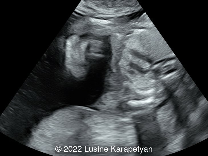
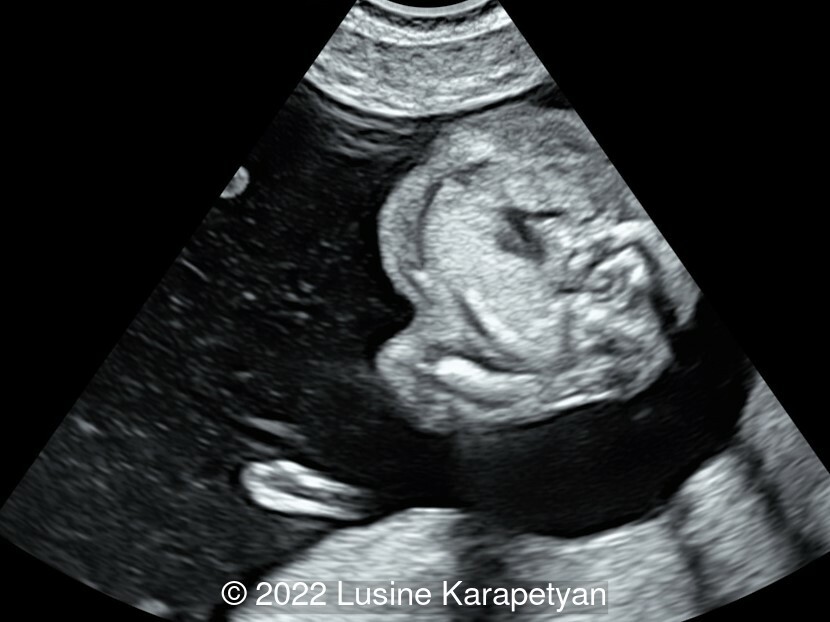
Case Report: 27-year-old G2P1 was referred to our unit for first trimesteric screening. A cardiac anomaly was detected, which was confirmed at 15 weeks. Images and videos performed at 15 weeks are presented. No other anomaly was detected.
View the Answer Hide the Answer
Answer
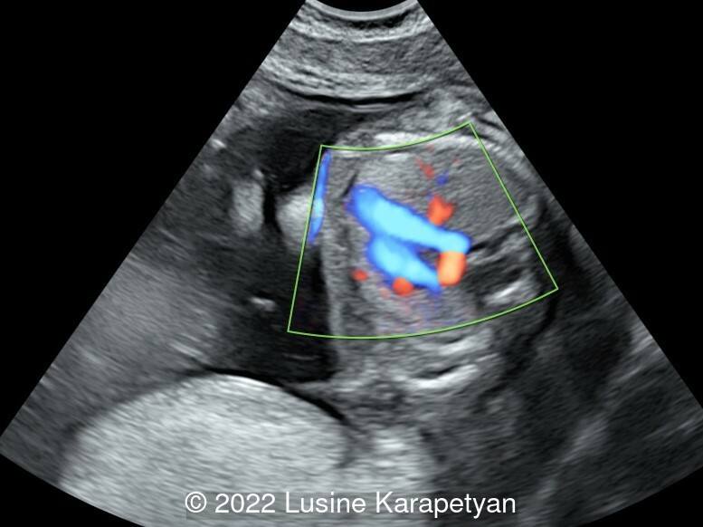
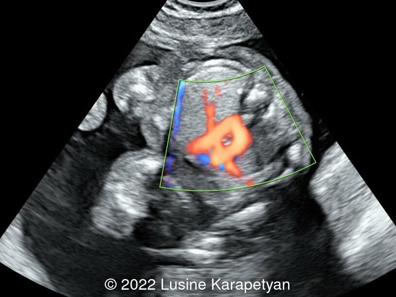
We present a case of double aortic arch. The initial fetal echocardiogram revealed normal intracardiac anatomy, and a double aortic arch with antegrade flow in both arches.
Discussion
Fetal double aortic arch (DAA) is a rare congenital heart disease affecting approximately 0.005% of fetuses. A double aortic arch is a congenital anomaly associated with the formation of a vascular ring, which can cause varying degrees of airway compression. This can be subclinical or clinical, manifesting as acute stridor, severe respiratory compromise or symptoms of chronic airway compression.
The diagnosis of double aortic arch on prenatal ultrasound is made when the ascending aorta appears to divide into two branches coursing on either side of the trachea. In 70% of double aortic arch cases, the right arch is dominant, but antegrade flow is commonly demonstrated in both aortic arches. The presence of a right aortic arch should prompt more careful evaluation to exclude a double aortic arch. The left aortic arch is often smaller than the right aortic arch, such that visualization of the diminutive left aortic arch is poor and can be overlooked on prenatal imaging [1].
The three-vessel tracheal view is the most characteristic view for double aortic arch diagnosis, but it alone is not enough. When an arch shows atresia, a vascular ring is not always typical due to an interruption in the blood flow of the atretic segment. The differential diagnosis with a right aortic arch then becomes challenging. Therefore, multiple views should be performed to better observe the origin, course, and branches of the two arches. The following steps and key points for systematic examination are recommended:
- Check for the presence of a the right aortic arch on the three vessel tracheal view.
- Search for the bifurcation of the ascending aorta in the left ventricular outflow tract view; trace the ascending aorta into the arch as the bifurcation is often not at the origin of the ascending aorta.
- Use the three-vessel tracheal view and sagittal view of the aortic arch to confirm that the left and right arches arise from the ascending aorta and are connected to the descending aorta.
- The three vessel trachea view is used to identify a complete O-shaped vascular ring formed by the left and right arches surrounding the trachea and esophagus. Because these two arches may not be at the same level, the probe may need to be slightly tilted.
- Measure the inner diameter of the left and right arches to determine the type of double aortic arch.
- Confirm the branching patterns of the left and right aortic arches on the sagittal views and coronal views of the aortic arch.
- Ascertain the presence of associated intracardiac or extra-cardiac malformations with careful examination of the heart and other systems, including the thymus.
- Consider obtaining chromosome and gene tests when necessary.
- In order to better display a small non dominant arch, reduce the scale of blood flow velocity in the color Doppler mode. It can be difficult to identify the fibrous cord formed by a partial atresia of the nondominant arch, therefore the vascular ring can be diagnosed indirectly by identifying the blind end or diverticulum of the arch. When the left arch is small and difficult to visualize, the right arch should not be mistaken for the right pulmonary artery, which would be travelling in the same direction [2].
Postnatal assessment should comprise echocardiography and cross-sectional imaging in the form of cardiac MRI/computed tomography. Bronchoscopy may be considered to exclude subclinical airway compression.
References
[1] Van Poppel MPM, Pushparajah K, Lloyd DFA, et al. Insights from fetal cardiac magnetic resonance imaging in double aortic arch. Ultrasound Obstet Gynecol. 2020 Oct;56(4):636-639.
[2] Guo Q, Kong Y, Zeng S et al. Fetal double aortic arch: prenatal sonographic and postnatal computed tomography angiography features, associated abnormalities and clinical outcomes. BMC Pregnancy and Childbirth. 2020;20:614.
Discussion Board
Winners

Dianna Heidinger United States Sonographer

Javier Cortejoso Spain Physician

Pawel Swietlicki Poland Physician

Fatih ULUC Turkey Physician
Umber Agarwal United Kingdom Maternal Fetal Medicine

Igor Yarchuk United States Sonographer
belen garrido Spain Physician

Andrii Averianov Ukraine Physician

Ana Ferrero Spain Physician

Alexandr Krasnov Ukraine Physician

Mayank Chowdhury India Physician
filiz halici öztürk Turkey Physician
Omayyah Dar Odeh Jordan Physician

Vladimir Lemaire United States Physician

Shilpen Gondalia India Physician

Ivan Ivanov Russian Federation Physician

Tatiana Koipish Belarus Physician

Halil Mesut Turkey Physician

Rushina Patel United States Sonographer

Caroline Reichert Garcia Brazil Physician

Anita Silber Israel Physician
Olivia Ionescu United Kingdom Physician

Rebecca Evans Australia Sonographer

Suat İnce Turkey Physician

Muradiye Yıldırım Turkey Physician

Dimitrios Spiliopoulos Greece Physician

Ta Son Vo Viet Nam Physician

Deval Shah India Physician

Murat Cagan Turkey Physician
gholamreza azizi Iran, Islamic Republic of Physician

Ionut Valcea Romania Physician
Karin Tinnemeier Germany Physician

TEJAS TAMHANE India Physician
Luis Goncalves United States Physician
Borisova Elena Russian Federation Physician

Jose Sanin Colombia Physician
ÇİĞDEM KUNT İŞGÜDER United States
Yasmeen Khalil United States
Soumya P United States

Seda Cam Turkey Physician

Kathrine Montagne United States Sonographer