Case of the Week #557
(1) High Risk Pregnancy Doctors, Frisco, Texas, United States of America; (2) St Mary's Medical Center, San Francisco, CA, United States
Posting Dates: April 15 - April 30, 2022
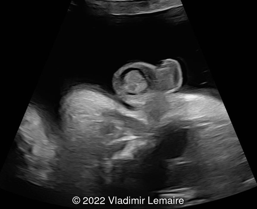
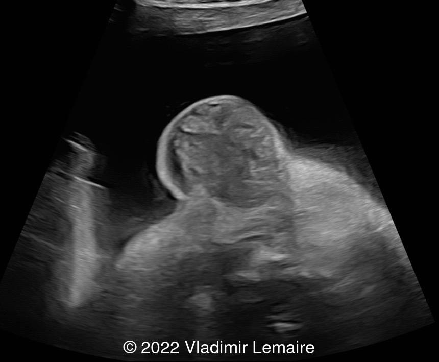
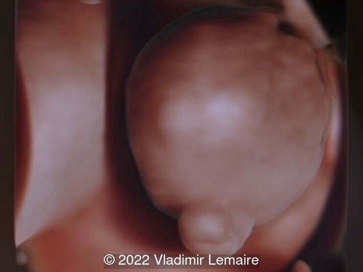
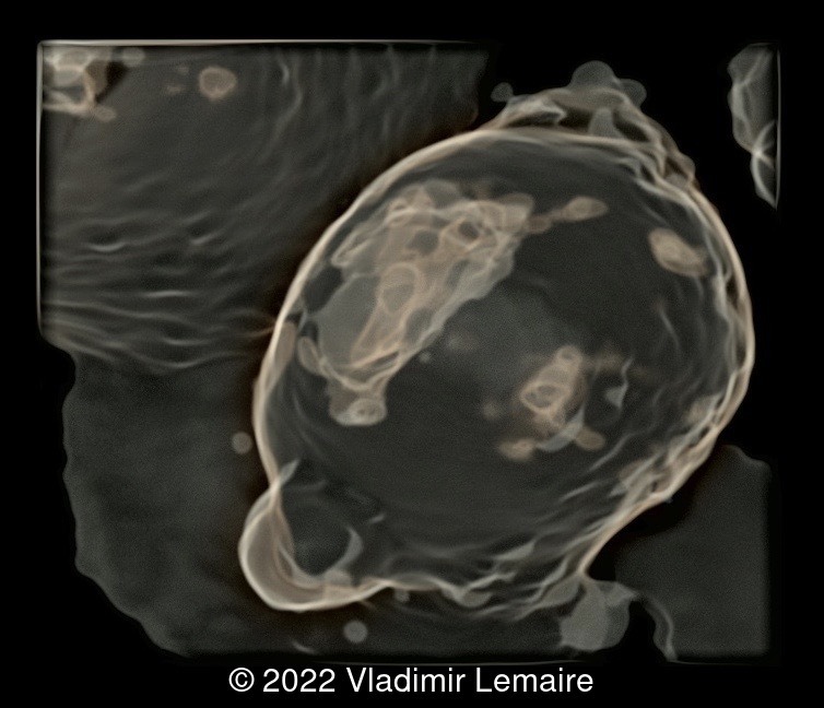
Case Report: A 40-year-old patient, G1P0, presented to our office for a biophysical profile at 34 w + 5 d with the following findings:
View the Answer Hide the Answer
Answer
We present a case of inguinoscrotal hernia. Ultrasound imaging revealed an enlarged fetal scrotum with a complex mass in the left side of the scrotum, which did not show increased blood flow with color Doppler. The contralateral testicle appeared normal. Although peristaltic movements were not documented during our evaluation, our diagnosis was based on the appearance of the mass and the absence of increased blood flow with color Doppler. Our diagnosis was confirmed after birth.
Discussion
Inguinal hernia is found in approximately 1.5% of neonates and is more common in males [1]. In premature infants weighing <1500g, the incidence of inguinal hernia is increased to 11% [2]. It is rarely diagnosed prenatally with less than 15 cases reported in the literature [3-5].
The processus vaginalis is present in the developing fetus at 12 weeks gestation. The testes descend at 7 to 8 months of gestation through the inguinal canal and into the scrotum. The obliteration of the processus vaginalis usually occurs by 2 years of age [6]. Inguinoscrotal hernia is commonly believed to result from increased intra-abdominal pressure [7], and an incomplete or abnormal obliteration of the processus vaginalis [6]. However, fetal inguinal hernias are extremely rare, and the intraabdominal pressure is similar or lower than the pressure in the amniotic cavity [8-10]. Amniotic pressure at 30 weeks has been measured to be 5 mm Hg [9] while intraperitoneal pressure measured during intrauterine transfusion in patients with Rh alloimmunization was measured at 2.5 mm Hg [10]. Ascites can increase abdominal pressure in utero [8], thus could be a causative factor in the development of inguinal hernia [11]. The etiology of prenatal inguinal hernia could also be due to an inherent weakness of the abdominal wall [12].
On ultrasound, inguinal hernia is characterized by a scrotal mass with smooth contour and predominantly solid content. Intestinal peristalsis can be seen [3,5,12]. Inguinoscrotal hernia has been associated with prematurity, especially infants weighing 500-1000g [2], fetal growth restriction [12,13], aneuploidy [12] and cystic fibrosis [14], but they are often isolated [3,5,15]. While the dreaded complication of intestinal strangulation is a concern, there are few cases reported in the literature with evidence of prenatal bowel dilatation concerning for obstruction [4,14]. Neither case reported intestinal ischemia; one was found to have anorectal malformation as a possible alternate explanation for the bowel dilation [4] and the other had cystic fibrosis [14].
Differential diagnosis for inguinal hernia includes hydrocele, testicular tumor such as teratoma, sacral meningomyelocele, hemangioma, testicular torsion, and meconium peritonitis. A hydrocele presents with a fluid-filled space surrounding testis and often has a “half moon” appearance [16]. A teratoma appears as complex heterogeneous mass with solid and cystic components, though more commonly occurs in an undescended testicle and presents as an abdominal mass in utero [17]. Testicular torsion is diagnosed by an enlarged testis and epididymis with accumulation of hemorrhagic fluid between the visceral and parietal layers of the tunica vaginalis and outside the tunica vaginalis resulting in a “double-ring hemorrhage.” Additionally, there is absence of testicular flow on color Doppler [18].
References
[1] Chang SJ, Chen JYC, Hsu CK. The incidence of inguinal hernia and associated risk factors of incarceration in pediatric inguinal hernia: a nation-wide longitudinal population-based study. Hernia. 2016 Aug;20(4):559-63.
[2] Kumar VHS, Clive J, Rosenkrantz TS, et al. "Inguinal hernia in preterm infants (< or = 32-week gestation)." Pediatr Surg Int. 2002 Mar;18(2-3):147-52. 3. Khatib N, Goldstein I, Vitner D, et al. "Prenatal diagnosis of scrotal-inguinal hernia: two case reports and review of the English literature." Eur J Obstet Gynecol Reprod Biol. 2013 Nov;171(1):9-11.
[4] Ronzoni S, Melamed N, Kingdom JC, et al. "Prenatal diagnosis of inguinoscrotal hernia associated with bowel dilatation: a pathogenetic hypothesis." Prenat Diagn. 2015 Nov;35(11):1151-3.
[5] Massaro G, Sglavo G, Cavallaro A, et al. "Ultrasound prenatal diagnosis of inguinal scrotal hernia and contralateral hydrocele." Case Rep Obstet Gynecol. 2013;2013:764579.
[6] Rowe MI, Copelson LW, Clatworthy HW. "The patent processus vaginalis and the inguinal hernia." J Pediatr Surg. 1969 Feb;4(1):102-7.
[7] Light HG, Routledge JA. "Intra-Abdominal Pressure Factor in Hernia Disease." Arch Surg. 1965 Jan;90:115-7.
[8] Katsura D, Takahashi Y, Iwagaki S et al. "Changes in Intra-Amniotic, Fetal Intrathoracic, and Intraperitoneal Pressures with Uterine Contraction: A Report of Three Cases." Case Rep Obstet Gynecol. 2018; 2018: 4281528. Published online 2018 Sep 12.
[9] Sideris IG, Nicolaides KH. "Amniotic Fluid Pressure during Pregnancy." Fetal Diagn Ther 1990;5:104–108.
[10] Nicolini U, Talbert DG, Fisk NM et al. "Pathophysiology of pressure changes during intrauterine transfusion." Am J Obstet Gynecol. 1989 May;160(5 Pt 1):1139-45.
[11] Yankes JR, Bowie JD, Effmann EL, et al. "Antenatal diagnosis of meconium peritonitis with inguinal hernias by ultrasonography. Therapeutic implications." J Ultrasound Med. 1988 Apr;7(4):221-3
[12] Paladini D, Palmieri S, Morelli PM, et al. "Fetal inguinoscrotal hernia: prenatal ultrasound diagnosis and pathogenetic evaluation." Ultrasound Obstet Gynecol. 1996 Feb;7(2):145-6.
[13] Kesby G, Beilby R, Petroni M. "Fetal inguinoscrotal hernia: sonographic diagnosis and obstetric management." Ultrasound Obstet Gynecol. 1997 Nov;10(5):359-61.
[14] Allen LM, Nosovitch JT, Silverman RK, et al. "Prenatal diagnosis of an inguinoscrotal hernia in a fetus with cystic fibrosis." J Ultrasound Med. 2004 Oct;23(10):1391-4.
[15] Meizner I, Levy A, Katz M, et al. "Prenatal ultrasonographic diagnosis of fetal scrotal inguinal hernia." Am J Obstet Gynecol. 1992 Mar;166(3):907-9.
[16] Pretorius DH, Halsted MJ, Abels W, et al. "Hydroceles identified prenatally: common physiologic phenomenonti" J Ultrasound Med. 1998 Jan;17(1):49-52.
[17] Siu SS, Leung TN, Leung TY, et al. "Prenatal diagnosis of intra-abdominal mature testicular teratoma." J Ultrasound Med. 2001 Nov;20(11):1257-60.
[18] Herman A, Schvimer M, Tovbin J et al. "Antenatal sonographic diagnosis of testicular torsion." Ultrasound Obstet Gynecol. 2002 Nov;20(5):522-4.
Discussion Board
Winners

Albert Buwono Indonesia Physician

Dianna Heidinger United States Sonographer

PEDRO MORALES UTRILLA Spain Physician

Javier Cortejoso Spain Physician

Fatih ULUC Turkey Physician

Kristína Bihariová Slovakia Physician

Umber Agarwal United Kingdom Maternal Fetal Medicine
Cem Sanhal Turkey Physician
Andrii Averianov Ukraine Physician

Ana Ferrero Spain Physician

Alexandr Krasnov Ukraine Physician

Adrian Popa Romania Physician
Omayyah Dar Odeh Jordan Physician
silvio tartaglia Italy Physician

Oskar Sylwestrzak Poland Physician

Shilpen Gondalia India Physician
Ivan Ivanov Russian Federation Physician

Sara Abdallah Salem Egypt Physician
Halil Mesut Turkey Physician

Rushina Patel United States Sonographer
Anita Silber Israel Physician

lan nguyen xuan Viet Nam Physician
Shafiga Hamzayeva Azerbaijan Physician

Andrii Telytchenko Ukraine Physician
Suat İnce Turkey Physician
Victoria Giang Viet Nam Physician
Liem Dang Le Viet Nam Physician

Amparo Gimeno Spain Physician
Yasemin Dogan Turkey Physician
Tatiana Shpilevaya Russian Federation Physician
Ta Son Vo Viet Nam Physician

ALBANA CEREKJA Italy Physician
SAMUEL GELVEZ TELLEZ Colombia Physician
Murat Cagan Turkey Physician

Sonio Sonio France AI
Charlotte Conturie United States Physician
Ionut Valcea Romania Physician
Vu Hung Viet Nam Physician
reyhan ayaz Turkey Physician
Chantal Mayer United States
Vera Aladinskaya United States
JUAN PEDRO ALVARADO DAVILA Mexico Physician

Đặng Mai Quỳnh Viet Nam Physician