Normal appearance of Brain Vesicles: week by week

Dr Vladimir Lemaire, Dr Violetta Lozovyy
High Risk Pregnancy Doctors, Frisco, Texas, United States of America

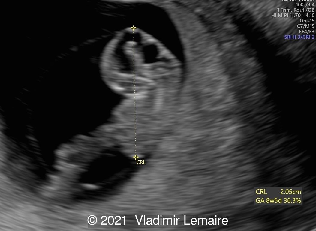
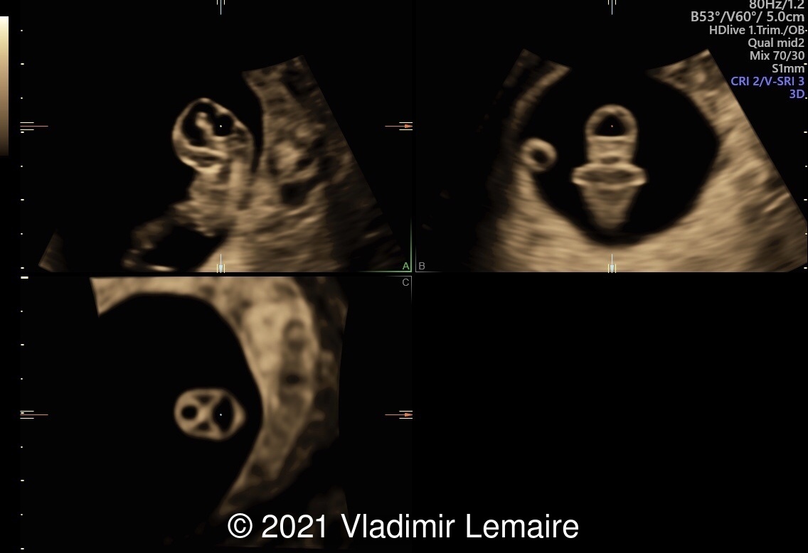
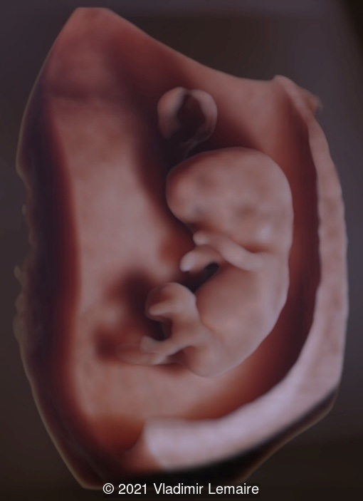
The following images demonstrate the normal appearance of the brain vesicles at 8 weeks of gestation.

The cephalic end of the neural tube initially contains three vesicles: the prosencephalon (forebrain), the mesencephalon (midbrain), and the telencephalon (hindbrain). The prosencephalon later differentiates into the telencephalon and diencephalon, the rhombencephalon divides into the metencephalon and myelencephalon, while the mesencephalon remains unchanged.

2D ultrasound showing early development of the brain.
Image 1 2D ultrasound showing early development of the brain.
Three-dimensional ultrasound in multiplanar display of an embryo at 8weeks of gestation showing the brain vesicles.
Image 2 Three-dimensional ultrasound in multiplanar display of an embryo at 8weeks of gestation showing the brain vesicles.
A rendered image of the same embryo in Silhouette mode.
Image 3 A rendered image of the same embryo in Silhouette mode.
Image obtained by augmenting the transparency effect and adjusting light effects.
Image 4 Image obtained by augmenting the transparency effect and adjusting light effects.

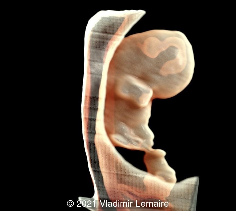
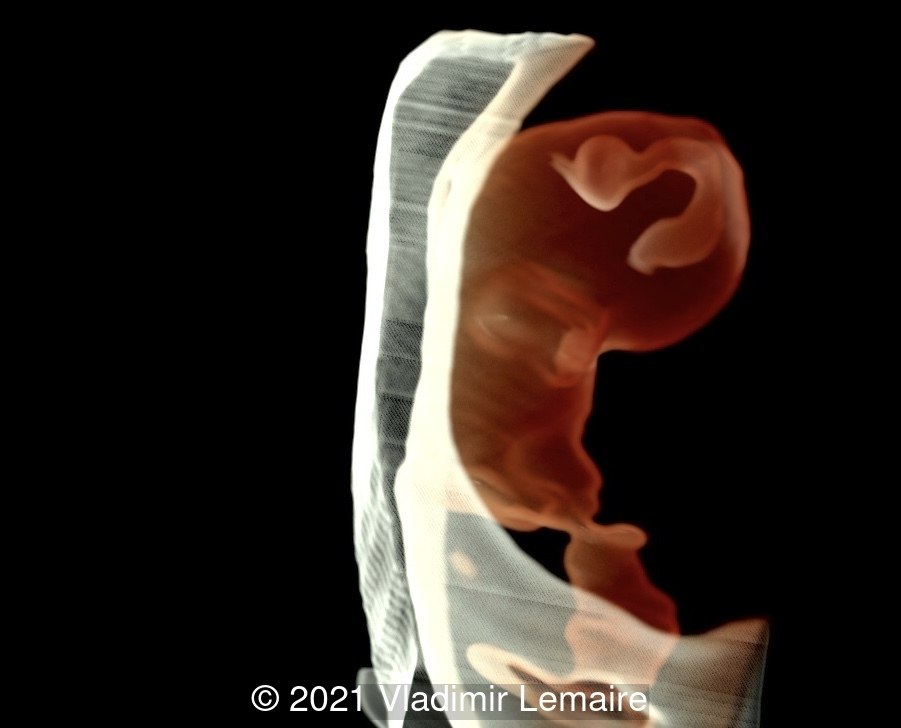
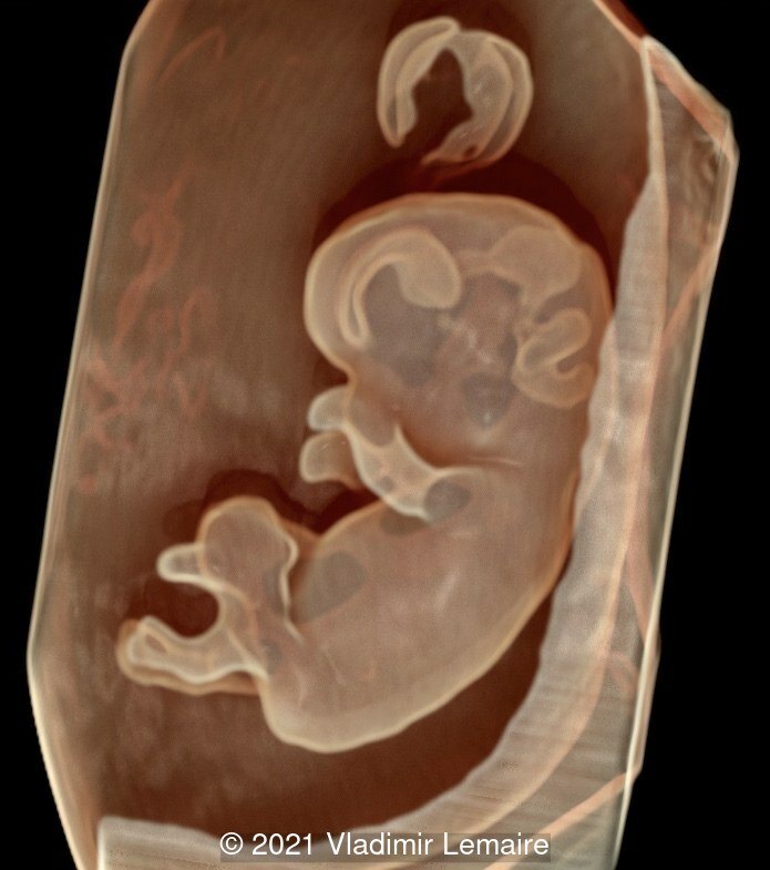
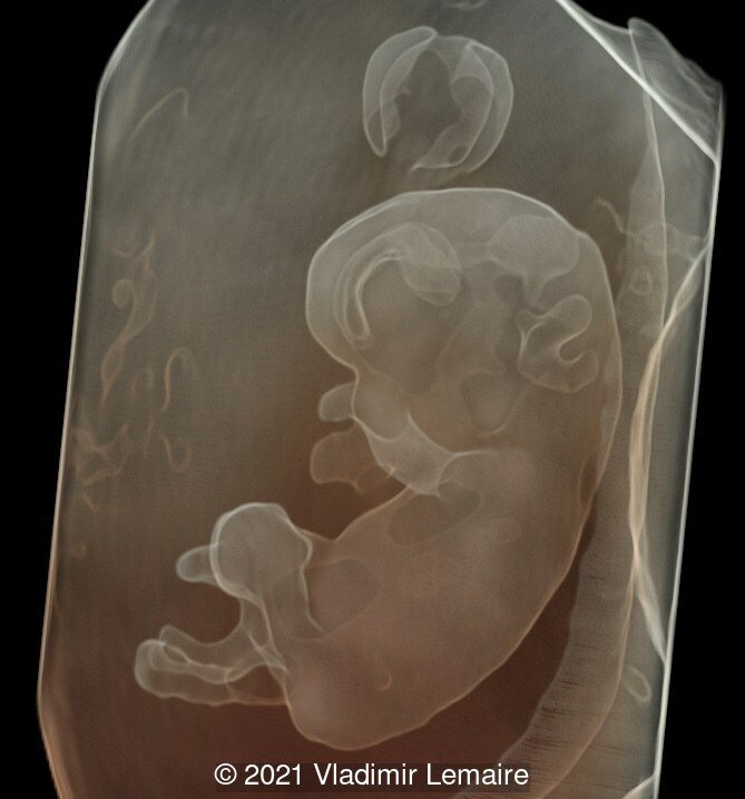
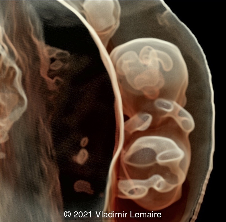
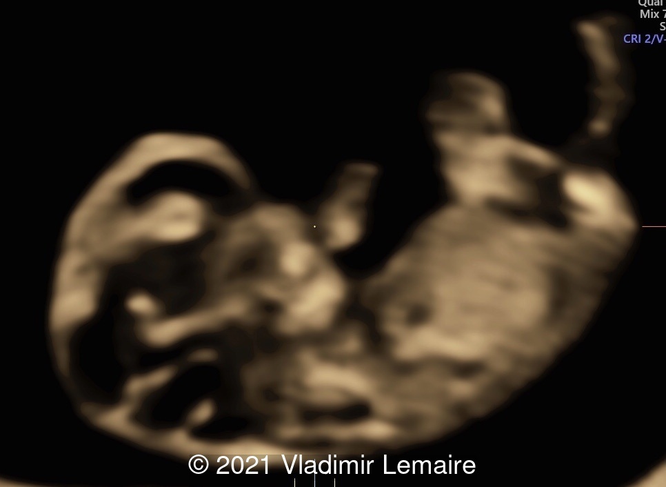
The following images demonstrate the normal appearance of the brain vesicles at 9 weeks of gestation.  At this point are easily identifiable: the telencephalon vesicles, the diencephalon, and the rhombencephalon.

3D image of a normal embryo with a crown-rump length of 30.8 mm. Surface rendering with shadow highlights the outer surface of the embryo, umbilical cord and yolk sac.
Image 5 3D image of a normal embryo with a crown-rump length of 30.8 mm. Surface rendering with shadow highlights the outer surface of the embryo, umbilical cord and yolk sac.
Lateral view of early brain development using Silhouette mode.
Image 6 Lateral view of early brain development using Silhouette mode.
Image obtained by increasing the transparency effect and adjusting light effects.
Image 7 Image obtained by increasing the transparency effect and adjusting light effects.
Frontal view of early brain development at 9 weeks (+ 6 days) of gestation. Bilateral telencephalon, midbrain and rhombencephalon are depicted.
Image 8 Frontal view of early brain development at 9 weeks (+ 6 days) of gestation. Bilateral telencephalon, midbrain and rhombencephalon are depicted.
Demonstration of early brain development using Volume Contrast Imaging (VCI) with 1-2 mm thickness. Note the development of the fourth ventricle at this gestational age. The choroid plexus of the fourth ventricle is also visible.
Image 9 Demonstration of early brain development using Volume Contrast Imaging (VCI) with 1-2 mm thickness. Note the development of the fourth ventricle at this gestational age. The choroid plexus of the fourth ventricle is also visible.

References:

Pooh RK. "Recent advances in 3D ultrasound, silhouette ultrasound, and sonoangiogram in fetal neurology." Donald School J Ultrasound Obstet Gynecol 2016;10(2):193-200.

Bault, JP, et al. "Echo-Anatomie de l'Embryon (7-10 sa)." Echo-Anatomie Normale du Foetus. Montpellier: Sauramps Medical; 2021. pgs 16-17.

Abuhamad A, et al. "The fetal central nervous system." First trimester ultrasound diagnosis of fetal abnormalities. Philadelphia: Wolters Kluwer; 2018. pgs 113-115.

Discussion Board

Start a discussion about this article
Add bookmark Bookmarked

Menu